J.ophthalmol.(Ukraine).2022;2:68-72.
|
http://doi.org/10.31288/oftalmolzh202226872 Published on-line: 30 April 2022 On the 75th anniversary of the establishment of Ophthalmic Pathomorphology Laboratory at the Filatov Institute of Eye Disease and Tissue Therapy E. V. Maltsev SI "The Filatov Institute of Eye Diseases and Tissue Therapy of the NAMS of Ukraine"; Odesa (Ukraine) TO CITE THIS ARTICLE: Maltsev EV. On the 75th anniversary of the establishment of Ophthalmic Pathomorphology Laboratory at the Filatov Institute of Eye Disease and Tissue Therapy. J.ophthalmol.(Ukraine).2022;2:68-72. http://doi.org/10.31288/oftalmolzh202226872 The article focuses on the results of diagnostic studies and research performed in the Ophthalmic Pathomorphology Laboratory over the 75 years of its existence. More than 250,000 histological studies of surgical clinical material and biopsy material from more than 100,000 patients have been performed over the whole period of existence of the laboratory since 1946. In addition, about 1000 science works including 26 monographs have been published, and dozens of ophthalmologists were provided special assistance in performing morphological studies for their doctoral dissertations and candidate of science theses.
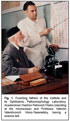
Ophthalmic pathomorphology is the sub-specialty of ophthalmology that focuses on the structural changes in the eye of humans or laboratory animals with different diseases. The Morphology Research Laboratory at Odesa Institute of Eyes Diseases was established soon after the Great Patriotic War, in 1946. Valentin Valentinovich Voino-Yasenetsky, a founder of the laboratory, had moved to Odesa by invitation of Academician V.P. Filatov, Director of the institute. This did not happen all of a sudden. The point is that the director was sure that the new institute staff member was a person of high moral standards because V.P. Filatov knew what kind of person the newcomer’s father was. Valentin Feliksovich Voino-Yasenetsky, the father of Valentin Valentinovich, was an outstanding Russian and Soviet surgeon (a general surgeon who had performed numerous kinds of surgeries on different systems and organs), Doctor of Medical Science, and professor. In addition, he was the world-famous author of Essays on the Surgery of Pyogenic Infections and other works, for which he was granted the Stalin Prize of the first degree in 1946. It is noteworthy that he also was Bishop of Tashkent and Turkestan from 1923 to 1927 (a period of murderous persecution of church), and Archbishop of Simferopol and Crimea from 1946 to 1961. He was arrested three times and spent years in internal exile over his life. On November 22, 1995, he was canonized by the Ukrainian orthodox Church for his piety. Therefore, it had been easy to guess that any child of this man could not be brought up wrongly by his/her father, which was subsequently confirmed by life itself. The Ophthalmic Pathomorphology Laboratory founded by would-be professor V.V. Voino-Yasenetsky has changed its name several times over the years, but the essentials remained the same. The laboratory has been focused first of all on (a) studies of the clinical material received from the departments of the institute and from all over Ukraine and Moldavia and, to a less extent, on (b) scientific research (Fig. 1).
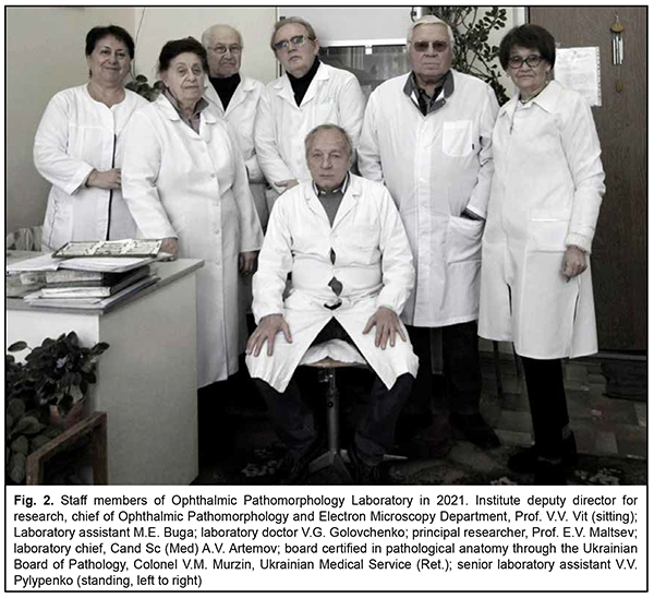
This is understandable, because the clinical material for histological studies is most commonly received from cancer department and pediatric department of the institute, and adult patients and parents of pediatric patients are always looking forwards for the results of these studies. More than 250,000 histological studies of surgical clinical material and biopsy material from more than 100,000 patients have been performed over the whole period of existence of the laboratory since 1946. That is, almost a sixth of patients of the polyclinic and treatment departments of the institute have become patients of the laboratory. The percentage is even higher for patients of the in-patient unit of the institute. Patients of eye cancer department account for as much as 90%, while the pediatric department, trauma and burn departments, cornea disease department and endovitreal surgery department account for the rest of the surgical clinical material and biopsy material obtained from the departments of the institute (which is completely understandable). All these studies are conducted by the staff of the laboratory (Fig. 2) headed by Professor V.V. Vit, who succeeded his teacher, Professor V.V. Voino-Yasenetsky, when the latter retired.
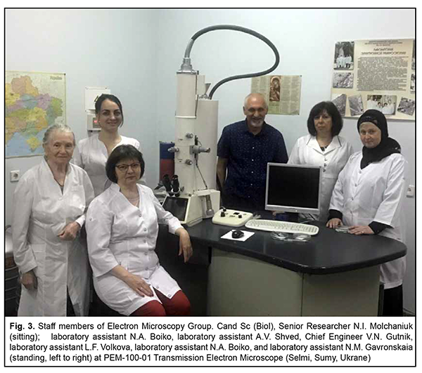
Electron Microscopy Group is another subdivision of the Ophthalmic Pathomorphology Department of the institute, and was established in 1968 under the instruction of and with the active participation of the director of the institute, Academician N.A. Puchkovskaia. The group was initially headed by Cand Sc (Med) N.Ie. Dumbrova, who later became doctor of medical science and professor, and was heading the laboratory until she passed away in 2015. She was succeeded by Cand Sc (Biol), Senior Researcher Natalia Ivanovna Molchaniuk, who has been working in the group since 1989. Fig. 3 shows the staff members of the group.
In our opinion, the major scientific achievements of the laboratory in the reported period are the following. The histomorphological results of keratoplasty using cadaver grafts preserved by the method of V.P. Filatov have been described and reviewed. A technique of intralamellar keratoplasty has been for the first time proposed. Foundations have been laid for studies of immune responses to transplantation of organs and organ parts. The damaging effects of laser and ultrasound on ocular structures have been have been identified and described. Ultrastractural changes in ocular tissues due to the effects of laser, ultrasound, electromagnetic pulse, and low doses of ion radiation have been for the first time described and reviewed. It has been demonstrated that therapeutic cataract treatment should be differential and not universal as it is in current eye care practice. It has been demonstrated that oncogenic factors such as cancerogenic chemicals, ionizing radiation, and chronic damage to the epithelial cells of the lens did not result in the development of lens malignancies, possibly due to some still unknown properties of the epithelial cells of the lens. It has been found that a uveal nevus can be distinguished from a uveal melanoma, irrespective of its size and pigmentation. It has been demonstrated that tumor cell anaplasia does not depend on melanocytic proliferation (i.e., there is no increase in tumor malignancy grade). It has been found that specific immune response plays an important role in the implementation of the therapeutic effect of various organ preserving treatment techniques (photocoagulation, brachytherapy). New data on mushroom-shaped choroidal tumors has been obtained and a new histogenetic concept for intraocular melanomas proposed. It has been established for the first time that dithizone-induced diabetes mellitus in rabbits is an optimal model for investigating the neurodegenerative processes developing in the retina in diabetic retinopathy. A rabbit model of pterygium has been proposed for the first time and the methodology of pterygium removal using electric welding of biological tissues has been developed. The potential of pharmacological correction of toxic injury to the optic nerve using aloe vera and RNA hydrolysate has been demonstrated histochemically and ultrastructurally in animals. Sixteen doctors of medical science and 60 candidates of medical science have been either trained or provided special assistance in performing their studies, about 1000 articles and other science works have been published in journals and other publications, and 26 monographs have been published over the whole period of existence of the laboratory since 1946. Therefore, every three years a new book written by a laboratory staff member either solely or in co-authorship has been published. We would like to conclude this article with a list of the 26 monographs (laboratory staff members are working on three more monographs). Artemov AV. [Non-toxic cancer therapy: pathophysiological mechanisms and clinical features]. Kharkiv: Reader Publishing House; 2003. Russian Artemov AV. [Donor cornea in current disease patterns]. Odesa: Interprint; 2007. Russian Artemov AV. [Intraocular mushroom-shaped tumors: a new histogenetic concept for intraocular melanoma]. Odesa: Interprint; 2008. Russian Artemov AV. [A universal concept of aging: an unexpected look at the problem through corneal epithelium]. Odesa: Interprint; 2008. Russian Artemov AV, co-author. [Herbal therapy for cancer, infection and general medical conditions: experience and recommendations in schemes, methodologies and formulations]. Kharkiv; 2010. Russian Artemov AV. [General pathology: a critical review of basic components]. Odesa: Interprint; 2011. Russian Artemov AV. [Issues of transplantation donorship: the law and constitutional guarantees]. Odesa: Interprint; 2014. Russian Artemov AV. [Aging: inevitability as a consequence of coincidence]. Odesa: Interprint; 2018. Russian Bobrova NF, Vit VV. [Atlas of congenital and acquired eye disorders]. Odesa; 2006. Russian Vit VV. [Structure of the human visual system]. Odesa: Astroprint; 2003. Russian Vit VV. [Tumor disease of the eye. In 2 volumes]. Odesa: Astroprint; 2009. Russian Vit VV. [Disorders of the eye, ocular adnexa and orbit. In 2 volumes]. Odesa: Astroprint; 2019. Russian Bobrova NF, editor. Pasyechnikova NV, Naumenko VO, Vit VV, Sorochinska TA. [Retinoblastoma]. Odesa: Izdatelskii Dom; 2020. Russian Voino-Yasenetsky VV. [Histoincompatibility and ways for overcoming it]. Moscow: Meditsina; 1965. Russian Puchkovskaia NA, Barkhash SA, Bushmich DG, Voino-Yasenetsky VV, Muchnik SR. [Basics of corneal transplantation]. Kyiv: Zdorov’ie; 1971. Russian. Puchkovskaia NA, editor. Barkhash SA, Voino-Yasenetsky VV, Marmur RK, et al. [Tumors of the eye, ocular adnexa and orbit]. Kyiv: Zdorov’ie; 1978. Russian Voino-Yasenetsky VV. [Ocular tissue growth and changes in eye disease and trauma]. Kyiv: Vyshcha shkola; 1979. Russian Puchkovskaia NA, editor. Voino-Yasenetsky VV, Gorgiladze TU, Kashintseva LT, et al. [Atlas of eye diseases]. Moscow: Meditsina; 1981. Russian. Puchkovskaia NA, Voino-Yasenetsky VV. [Secondary dystrophic and structural changes in the anterior segment of the eye]. Moscow: Meditsina; 1985. Russian Maltsev EV. [Lens]. Moscow: Meditsina; 1988. Russian Maltsev EV, Pavliuchenko KP. [Biological features and diseases of the lens]. Odesa: Astroprint; 2002. Russian Maltsev EV. [Methodology of scientific creativity in medicine]. Odesa: Astroprint; 2006. Russian Usov VIa, Maltsev EV. [Glasses, contact lenses, surgical correction of vision or informed choice]. Kyiv: Knyga Plus; 2006. Russian Maltsev EV, Zborovska OV, Dorokhova OE. [Fundamental aspects of the development and treatment of diabetic retinopathy]. Odesa: Astroprint; 2018. Russian Maltsev EV, Usov VIa, Krytsun NIu. [Pterygium – an ophthalmic enigma]. Odesa: Astroprint; 2020. Russian Maltsev EV. [Ophthalmic Pathomorphology in Ukraine]. Odesa: Astroprint; 2021. Russian. (In print) In addition, the results of our electron microscopy studies were heavily used in the following monographs: Bobrova NF, Skripnichenko ZM.[Toxic, congenital, and secondary cataracts]. Odessa: Feniks; 2017. Russian. Pasyechnikova NV, editor. [High-frequency welding of biological tissues in ophthalmology]. Odessa: Chornomor’ia; 2017. Ukrainian Pogrebnjak AD, Pogorielov M, Viter R, editors. Nanomaterials in Вiomedical Аpplication and Вiosensors.Springer Proceedings in Physics. Springer: Singapore, 2020; Volume 244. Bobrova NF, editor. Pasyechnikova NV, Naumenko VO, Vit VV, Sorochinska TA. [Retinoblastoma]. Odesa: Izdatelskii Dom; 2020. Russian
|