J.ophthalmol.(Ukraine).2016;2:37-40.
|
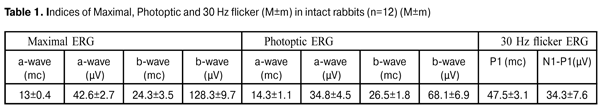
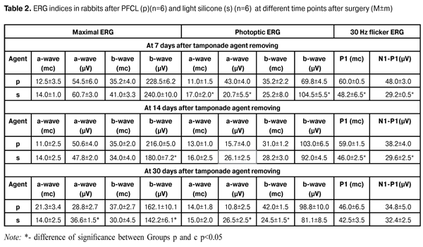
https://doi.org/10.31288/oftalmolzh201623740 Effect of two week tamponade using perfluorocarbon liquid and light silicone oil on the bioelectrical functional activity in the retina in rabbits D.V. Zhmuryk, Cand Sc (Med) 1 N.I. Khramenko, Cand Sc (Med) 2 S. B. Slobodyanik, Cand Sc (Med) 2 M.V. Miliienko, MD 1 1Kyiv Clinical Ophthalmology Hospital Eye Microsurgery Center 2Filatov Institute of Eye Diseases and Tissue Therapy, NAMS of Ukraine Kyiv, Odessa (Ukraine) E-mail: visus@ukr.net Introduction. Using perfluorocarbon liquid (PFCL) for short-term tamponade of the vitreous cavity expands the indications to surgical treatment of retinal detachment (RD) of various genesis. However, the question about affecting action of PFCL and about the maximal safe term of tamponade is still open. The purpose of the present paper was to determine the characteristics of the effect of two week PFCL tamponade on the bioelectrical functional activity in the rabbit’s retina and to compare the action of PFCL and light silicone oil (SO) with viscosity of 5 700 cSt in dynamics by electroretinography (ERG) at different time points after tamponade (7, 14 and 30 days). Material and Methods. The experiment involved 6 male rabbits (12 eyes). Each animal was performed vitrectomy following 14 day tamponade with PFCL (in the right eye) and light SO. ERG was performed at baseline as well as after tamponade removal at time points of 7, 14 and 30 days. Results. 14 day PFCL (perfluorodecalin) tamponade of the rabbit eyes induced similar bioelectric reaction in the retina, comparing to light SO tamponade (viscosity 5 700 sST). The reaction consisted of increased activity in photoreceptor and inner retinal layers at early terms and normalization of retinal functions at late terms (1 month) after tamponade removing with a decrease in inner layer reactivity. Conclusion. PFCL can be considered as a short-term tamponade agent. Key words: ultra structure of the retina, functional activity, electroretinography, perfluorocarbon liquids, silicone oil, experiment Introduction The modern stage in the development of surgical treatment for the posterior segment of the eye has been marked by new achievements. Since the beginning of the XXI century, vitreoretinal surgery has been developing rapidly: there have appeared small caliber surgery (23G, 25G, 27G), the latest generation of lasers, and optic devices to visualize clearly the ocular fundus during the surgery. It is constantly that physical and chemical features and biological inactivity of perfluorocarbon liquids (PFCL) are improved and more purified silicone oil (SO) and various vitreoretinal tools are developed. Perfluorocarbon liquids are an important tool in the vitreoretinal surgery, the general development of which is dependent on the development of each part. Using PFCL for short-term tamponade of the vitreous cavity extends indications for operative treatment of the retinal detachment (RD) of various geneses, and provides intra- and interoperation evacuation of the residual subretinal fluid and adequate retinal adaptation; PFCL can be used with a haemostatic purpose that enables to inject silicone oil (SO) on the intact retina and to perform additional laser coagulation (LC) [3, 5, 13]. Despite all this, the attitude of the vitreoretinal surgeons towards short-term tamponade of the vitreous cavity using perfluorocarbons (PFC) is ambiguous. The question of damaging action of PFC as well as of maximal safe tamponade duration is still open [10, 14, 15]. We have analyzed experimental papers on this issue and found that their results differ from each other. A part of researchers have reported on irreversible atrophic alterations after tamponade with duration of 48 hours [6], 2 weeks [4] and 1 month [1, 2, 13]. Other experimental studies have reported on the absence of the significant changes even after 1 month tamponade [7, 9]. However, they are difficult to compare since the experimental conditions are different, in particular, different techniques for removal of vitreous humor, various specific gravity of compounds used, and localization of the most damaging action not always taken into account. Saline solution has been always served as control. Thus, by reference of previous studies, it would be relevant to unify the conditions of our experiments: •to make experimental conditions most closely resembling real-life conditions (performing vitrectomy); •to use PFCL with high specific gravity to perform tamponade (in clinical practice, PFC used are of the same degree of purification and with various specific gravity: perfluorooctane (1.76 g/cm3), perfluorotributylamine (1.89 g/cm3), pfokalin (1.94 g/cm3) and the others) since the absence of damage when using them implies the safety of using PFCL with lower specific gravity; •to study alterations in the retina at different time points after tamponade; •to take into account localization of maximal damaging action; •to compare the effect on the retinal ultrastructure of PFCL with a standard tamponade agent, silicone oil (specific gravity 0.971-0.975 g/cm3). The purpose of the present paper was to determine the characteristics of the effect of two week PFC tamponade on the bioelectrical functional activity in the rabbit’s retina and to compare the action of PFC and light SO with viscosity of 5 700 cSt in dynamics performing electroretinography (ERG) at different time points after tamponade (7, 14 and 30 days). Material and Methods All interventions and euthanasia were performed in accordance with The International Guiding Principles for Biomedical Research Involving Animals developed by the Council for International Organizations of Medical Sciences [12]. The experiment involved 6 male chinchilla rabbits (12 eyes), weighted 3.5±0.5 kg and aged 6.5±0.5 months. The tamponade duration was 14 days. Each animal was performed ERG at baseline as well as time points of 7, 14 and 30 days after PFC tamponade of the vitreous cavity. In all cases the second eye (left one) served as control. In the control eyes tamponading agent was light SO with viscosity of 5 700 cSt (specific gravity<1.0 g/cm3). Surgical technique: Preoperatively, to anesthetize the experimental animals, 2mg/kg of thiopental sodium was injected intramuscularly and 0.5% of proparacaine was administered epibulbarly; mydriasis was achieved with epibulbar 1% atropine sulfate and 2.5% phenylephrine. Before surgery, 0.3% ofloxacin was used epibulbarly. 20G, 23G and 25G closed subtotal vitrectomy was performed using KFE-01 “MEDA-NN” Sonorus operating system (frequency up to 1 200 bpm; aspiration 150 mmHg). The vitrectomy was performed under the control of OPTONO pMi-8 operating microscope and Photon 2 light. 1.5 mL of PFCL perfluorodecalin (PFD) was administered in the vitreous cavity of the right eye. The cavity of the left eye was filled in with 1-1.5 mL of light SO with viscosity of 5 700 cSt. After vitrectomy, 0.3% ofloxacin ointment was put into the conjunctival cavity. The tamponade duration was 14 days. Tamponade was completed with preoperative operations described above. Removal of PFD was performed using KFE-01 “MEDA-NN” Sonorus operating system (aspiration 150 Mmhg) under the control of OPTONO pMi-8 operating microscope and Photon 2 light. Silicone oil was removed actively under the operating microscope control. ERG procedure Preoperatively, 0.5% proparacaine was administered epibulbarly; mydriasis was achieved with epibulbar 1% atropine sulfate and 2.5% phenylephrine. A full field (Ganzfeld) ERG examination was performed using electrophysiological test unit Retiscan according to ISCEV Standard ERG protocols. Scotoptic combined rod-cone response, photoptic cone response and photoptic cone response to 30 Hz flicker were analyzed. The contact lens electrode was placed on the cornea; the reference electrode was placed on the forehead skin at the medium line; and the ground electrode was placed to the ear skin. ERG was performed at time points of 7, 14 and 30 days after 14 day tamponade. The data on electrophysiology assay of the control (intact) animals are given in the Table 1. Results and Discussion At Day 7 after removal of a tamponading agent, rod and cone response to the flash in the inner layers (bipolar and Muller cells) in the peripheral retina didn’t differ in eyes with both PFCL and SO tamponade (Table 2) and was characterized not only by induced retinal activity but and decreased peak-time of the potentials in the inner layers. The former was expressed in an increase in a-wave and b-wave amplitude by 35% (р<0.05) and 82% (р<0.05), respectively, as compared to norm; the latter was decreased by 58% (р<0.05). Photoptic ERG revealed that the cone response in the photoreceptor layer was lower when using SO than when using PFCL as a tamponade agent; thus, a-wave amplitude was equal to 20.7±5.5 (µV) in the former and it was 2.1 times higher in the latter (р=0.001). Cone response to flash in the inner retinal layer was an increase in b-wave amplitude by 48% (р=0.001) in eyes with SO tamponade (Table 2).
30 Hz flicker ERG response reflects activity of the cone system to 30 Hz stimuli. That in eyes after PFCL tamponade was characterized by increased N1- Р1 (µV) wave amplitude by 65.5% (р=0.001) and longer peak time by 24.4% (р=0.03). 30 Hz flicker ERG didn’t differ from the norm in eyes after SO tamponade (Table 2). High frequency flicker ERG is considered to be a signal reflecting total resulting activity only of neuronal retinal elements not mediated by neuroglia since, according to Miller R.F., Dowling J.E., (1970) [11], glial Muller cells are unable to resolve flickers with a rate higher than 2-4 Hz. Thus, at Day 7 of follow up after tamponading agent removing, action of SO and PFCL resulted in almost similar increased response in photoreceptor and inner layers of the retina by 35-85% and delayed conduction in the inner layers (bipolar and Muller cells) by 58%. However, PFCL effect on the cones was more sparing as compared to that of light SO: SO tamponade resulted in a decrease in cone response, implicit time delay and too high response in the inner layers (by 48%). At Day 14 after tamponading agent removal, the inner retinal layers in the PFCL eyes responded to a flash with a higher b-wave amplitude than in eyes with light SO by 16.6% (р=0.02). So, it was equal to 216±5 (µV) (Table 2). Photoptic ERG didn’t differ significantly in groups; however, b-wave amplitude showed increased reactivity in the inner layers by 43% as compared to the norm (Table 1). 30 Hz flicker responses were recorded with a higher amplitude of N1- Р1 (µV) wave by 22 % (р=0.04); however, as compared to the norm, that didn’t alter significantly in both groups. Only latency was slowed in the neuronal elements of cones by 22% (р=0.001) to (59.0±1.5) mc in PFCL group. Thus, at Day 14 after tamponading agent removal, response was increased in the retinal inner layers (by 16.6% and more in PFCL) as well as in the inner layers of the cones, in both groups. At Day 30, Maximal ERG was actually within the normal range in SO group, only a-wave amplitude decreased by 14% (р=0/04) as compared to the norm. In PFCL group, a-wave amplitude was smaller in Maximal ERG and Photoptic ERG by 27% (р=0.006) and 59% (р=0.001), respectively, as compared to SO group; that was likely a reflection of early dystrophic alterations in the photoreceptors. Flash response of the inner cone layers was characterized by increased activity in both groups; herewith, b-wave amplitudes did not differ significantly (Table 2) and conduction delay was observed after PFCL removal. It should be noted that 30 Hz Flicker ERG was normalized in both groups. Thus, at early terms after tamponading agent (PFCL and SO) removal, rabbit’s retina respond was similar and characterized by actually equal increase in activity of photoreceptor and the inner retinal layers (supernormal ERG). At Day 14, response in the inner retinal layers remained increased in both groups, but with a less amplitude as compared with that at time point of 7 days (Table 2). At Day 30, in both groups, flash response in inner cone layers was still increased with more delayed conduction and decreased amplitude in photoreceptor response in PFCL group. The data on normalization of 30 Hz flicker ERG in both groups showed the safe ability of the retinal neurons to reproduce light rhythm. Conclusions As a result of the experiment, 14 day PFCL (perfluorodecalin) tamponade of the rabbit eyes induced similar bioelectric reaction in the retina, comparing to light SO tamponade (viscosity 5 700 sST). The reaction consisted of increased activity in photoreceptor and inner retinal layers at early terms and normalization of retinal functions at late terms (1 month) after tamponade removing with a decrease in inner layer reactivity. Therefore, PFCL can be considered as a short-term tamponade agent.
References
|