J.ophthalmol.(Ukraine).2020;6:61-63.
|
http://doi.org/10.31288/oftalmolzh202066163 Received: 14 August 2020; Published on-line: 21 December 2020 A case of radiation cataract found 29 years after radiation exposure N. V. Pasyechnikova1, P. A. Fedirko2, T. F. Babenko2 1 Filatov Institute of Eye Diseases and Tissue Therapy of the National Academy of Medical Sciences of Ukraine; Odesa (Ukraine) 2 Health Physics and Epidemiology Institute, National Research Center for Radiation Medicine, National Academy of Medical Sciences of Ukraine; Kyiv (Ukraine) E-mail: eye-rad@ukr.net TO CITE THIS ARTICLE: Pasyechnikova NV, Fedirko PA, Babenko TF. A case of radiation cataract found 29 years after radiation exposure. J.ophthalmol.(Ukraine).2020;6:61-3. http://doi.org/10.31288/oftalmolzh202066163 Background: Radiation cataracts are an acknowledged biological effect of radiation exposure. It has been demonstrated previously that, the mode value for the latent period for the identified post-Chornobyl cases of radiation cataract was 9 years. We present a case of radiation cataract with typical clinical features that manifested 29 years after radiation exposure. Material and Methods: A female patient, born in 1937, worked in the exclusion zone of the Chornobyl Nuclear Power Plant in early May of 1986; she is now under our regular supervision. Because over years, the patient had had the annual routine eye examination (including, but not limited to biomicroscopy, lens examination and red reflex photography using a fundus camera), the time of onset of specific lens opacity could be placed within a period of several months. Results: On examination performed on December 15, 2014, the right lens showed mild peripheral cortical opacity without signs of radiation cataract, and with vacuoli seen in the anterior subcapsular lens region. On examination performed on August 8, 2015, the right eye showed a mild, specific, posterior, central subcapsular opacification. Conclusion: We presented a case of radiation cataract, documented by fundus camera photography, with typical clinical features that manifested 29 years after radiation exposure. Detecting a radiation cataract so late after radiation exposure indicates that the changes in the eye exposed to radiation can be very durable. Keywords: cataract, radiation cataract, ionizing radiation, Chornobyl acciddent, latent period
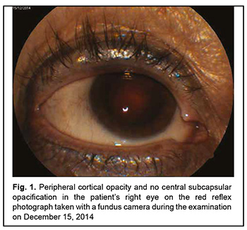
Introduction Findings of the post-Chornobyl eye examination studies in numerous cohorts of individuals exposed to radiation have changed our understanding of the effects of ionizing radiation on the eye. The new findings allowed us to change our view of radiation cataract, the most well known eye disease directly caused by radiation [1]. Since the disease has specific clinical features which are unique, post-Chornobyl cases of radiation cataract were easily distinguished and studied in detail [2-4]. The findings from the relevant studies have significantly changed our view of the ionizing radiation dose required to produce a radiation cataract [1, 3, 4]. Studies are underway on radiation cataractogenesis [5] and management of various retinal disorders [6, 7] that have been recently associated with ionizing radiation exposure [8-10]. The new data on the latent period, the time between radiation exposure and the appearance of lens opacities, is of importance. There is a lack of generalized data of the duration of the latent period, since in the past, most authors reported only on isolated cases of radiation cataract. Nevertheless, it has been generally recognized that radiation cataract may develop as early as a few years after radiation exposure [2]. We have demonstrated previously that, the mode value for the latent period for the identified post-Chornobyl cases of radiation cataract was 9 years [1, 3]. Case reports on late-onset radiation cataracts are important both from theoretical and practical points of view, because studying such cases is important for developing strategies for conservative and surgical management of survivors of radiation accidents and attacks, and helps to consider hypotheses on the pathogenesis of radiation cataract. Material and Methods A female patient born in 1937 is under our regular supervision. In early May of 1986, she was on a business trip to Chornobyl, visited the industrial site of the Chornobyl nuclear power plant (CNPP), and within a short period was at the direct line of sight from CNPP Unit 4. Although there is no direct individual dose measurement data available on the patient, bat reconstruction of individual doses by analytical and calculation methods may be performed, and we hope to report on the results in the future. After completing her business trip to Chornobyl in May 1986, the patient lived outside the areas of substantial radiation pollution [11-14], did not undergo radiological procedures, and was not involved in elimination of the consequences of any other radiation accident. Because over years, the patient had had the annual routine eye examination (visual acuity assessment and tonometry; in addition, biomicroscopy, lens examination and red reflex photography (using a fundus camera), ophthalmoscopy, and fundus photography (using a fundus camera) performed under dilated pupil conditions), the time of onset of specific lens opacity could be placed within a period of several months. Results Below are presented the results of eye examinations performed prior to and after finding signs of radiation cataract. On examination performed on December 15, 2014, no signs of radiation cataract were detected, but the lens showed mild peripheral cortical opacity (Fig. 1), and there was mild dry age-related macular degeneration (AMD). The fasting blood glucose level was 5.2 mmol/L.
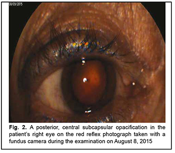
On examination performed on August 8, 2015, there was mild specific opacification in the central subcapsular region (Fig. 2). In addition, there were mild dry AMD and retinal angiopathy. The fasting blood glucose level was 5.1 mmol/L.
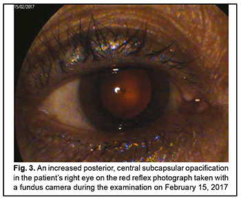
Thereafter, a rather fast progression of opacity was observed. Immediately after examination performed on February 15, 2017 (Fig. 3), because of progression of central subcapsular opacity in the right eye, the patient was recommended to have surgery.
Therefore, we presented a case of radiation cataract, documented by fundus camera photography, with typical clinical features that manifested 29 years after radiation exposure. Detecting a radiation cataract so late after radiation exposure indicates that the changes in the eye exposed to radiation can be very durable. We observed a relatively rapid decrease in transparency of the affected eye after specific lens opacity was found. These findings should be taken into account while (a) deciding whether an individual can be medically certified as capable of working under conditions of contact with sources of ionizing radiation, and (b) treating a job-related eye disease. References 1.Pasechnikova NV, Fedirko PA. Radiation cataract: new data received after Chornobyl accident. In: Proceedings of the International conference “Health effects of the Chornobyl accident – 30 years aftermath”. April 18-19, 2016. Kyiv, 2016. p.113. 2.Worgul BV, Medvedovsky C, Wu B. Use of non-subjective analysis of lens transparency in experimental radiation cataract research. Ophthalmic Res. 1995;27 Suppl 1:110-5. 3.Fedirko PA. [Radiation cataract as a late effect of the Chornobyl disaster]. Visnyk naukovykh doslidzhen. 2002;2:46-8. Ukrainian. 4.Fedirko PA, Babenko TF, Kolosynska OO, et al. Clinical types of cataracts in a long-term period after acute radiation sickness. Probl Radiac Med Radiobiol. 2019 Dec;24:493-502. 5.Zhou DD, Yao L, Guo KM, Lu CW. Cytogenetic evaluation of cataract patients occupationally exposed to ionizing radiation in northeast China. Genet Mol Res. 2016 Sep 16;15(3). 6.Korol AR, Zadorozhnyy OS, Naumenko VO, Kustryn TB, Pasyechnikova N.V. Intravitreal aflibercept for the treatment of choroidal neovascularization associated with pathologic myopia: A pilot study. Clin Ophthalmol. 2016 Nov 4;10:2223-2229. 7.Korol A, Kustryn T, Zadorozhnyy O, Pasyechnikova N., Kozak I. Comparison of efficacy of intravitreal ranibizumab and aflibercept in eyes with myopic choroidal neovascularization: 24-month follow-up. J Ocul Pharmacol Ther. 2020 Mar;36(2):122-125. 8.Babenko TF, Fedirko PA, Dorichevska RY, Denysenko NV, Samoteikina LA, Tyshchenko OP. The risk of macular degeneration development in persons antenatally irradiated as a result of Chornobyl NPP accident. Probl Radiac Med Radiobiol. 2016 Dec;21:172-7. 9.Yao X, Zhai M, Zhou L, Yang L. Protective effects of SND1 in retinal photoreceptor cell damage induced by ionizing radiation. Biochem Biophys Res Commun. 2019 Jun 30;514(3):919-925. 10.Mao XW, Boerma M, Rodriguez D, et al. Acute Effect of Low-Dose Space Radiation on Mouse Retina and Retinal Endothelial Cells. Radiat Res. 2018 Jul;190(1):45-52. 11.Vasylenko VV, Nechaev SY, Tsigankov MYa, et al. Results of comprehensive radiological - hygienic monitoring in some settlements of radiologically contaminated areas in Rivne region in 2017. Probl Radiac Med Radiobiol. 2018 Dec;23:139-152. 12.Vasylenko VV, Tsigankov MYa, Nechaev SYa, et al. Peculiarities of internal radiation doses due to 137Cs and 90Sr intake in population from Zhytomyr oblast in a late period after the Chornobyl NPP accident et al. Probl Radiac Med Radiobiol. 2013;(18):59-69. 13.Prylypko VA, Morozova MM, Petrychenko OO, Ozerova YY, Kotsubinskij OV. Morbidity rates in the NPP surveillance zone and radiologically contaminated areas. Probl Radiac Med Radiobiol. 2018 Dec;23:188-199. 14.Gunko NV, Korotkova NV. Variability of population gender and age composition in areas with the most intensive radiological contamination in Ukraine. Probl Radiac Med Radiobiol. 2018 Dec;23:153-163. The authors certify that they have no conflicts of interest in the subject matter or materials discussed in this manuscript.
|