J.ophthalmol.(Ukraine).2019;3:26-31.
|
Received: 22 March 2019; Published on-line: 27 June 2019 http://doi.org/10.31288/oftalmolzh201932631 Current antiseptics: a study on their antimicrobial activity and toxic effects on the corneal epithelium O.A. Nazarchuk, Cand Sc (Med); I.L. Chereshniuk, Cand Sc (Med); G.G. Nazarchuk, Cand Sc (Med); D.V. Palii, Cand Sc (Med) Pirogov Vinnytsia National Medical University; Vinnytsia (Ukraine) E-mail: shepelyuk.g.g@gmail.com TO CITE THIS ARTICLE: Nazarchuk OA, Chereshniuk IL, Palii DV. Current antiseptics: a study on their antimicrobial activity and toxic effects on the corneal epithelium. J.ophthalmol.(Ukraine).2019;3:26-31. http://doi.org/10.31288/oftalmolzh201932631 Background: Infectious agents can develop resistance to antibiotics, but antiseptics maintain their efficacy. It is still important to enhance our understanding of the effects of antiseptics on the macroorganism. Purpose: To investigate antimicrobial efficacy of the current antiseptic medications used in the ophthalmological practice, quaternary ammonium compounds (QACs) of decamethoxin (oftalmodek or OD) and miramistin (okomistin or OM), and to assess their effects on anterior corneal epithelial (ACE) DNA fragmentation and cell cycle in rats. Methods: We conducted a microbiological study to compare OD and OM for antimicrobial efficacy using serial two-fold dilutions. In addition, flow cytometry was used to investigate the effects of these medications on ACE cell apoptosis and proliferation in rats. Results: When compared with OM, OD demonstrated a higher antimicrobial efficacy against a wide spectrum of infectious agents (р < 0.001). Our in vivo flow cytometry study found no significant difference (p > 0.05) in fractions of ACE cells in various cell cycle phases between experimental eyes that received OD and intact fellow eyes. Experimental eyes in the okomistin group demonstrated depressed DNA synthesis in nuclei of ACE cells, with the fraction of ACE cells in the S phase that was 1.4-fold lower (р < 0.05) than in intact fellow eyes. In addition, the fraction of ACE cells in the G2+M phase and proliferative index of the ACE were 1.4-fold and 1.6-fold, respectively, lower (р < 0.05), whereas DNA fragmentation level in ACE cell nuclei was 1.5-fold higher (р < 0.05) than in intact fellow eyes. Conclusion: Oftalmodek has high antimicrobial activity against a broad spectrum of infectious agents and has no cytotoxic or proapoptotic effects. This was evidenced by the respective indices of its activity, which were 1.8 to 6.1-fold higher than those of okomistin (р < 0.001). However, the use of okomistin resulted in depressed DNA synthesis in nuclei of ACE cells, decreased proliferative index of the ACE, and increased DNA fragmentation level in epithelial cell nuclei compared to intact fellow eyes. Keywords: oftalmodek, okomistin, cornea, apoptosis, cytotoxicity, infection, antiseptics
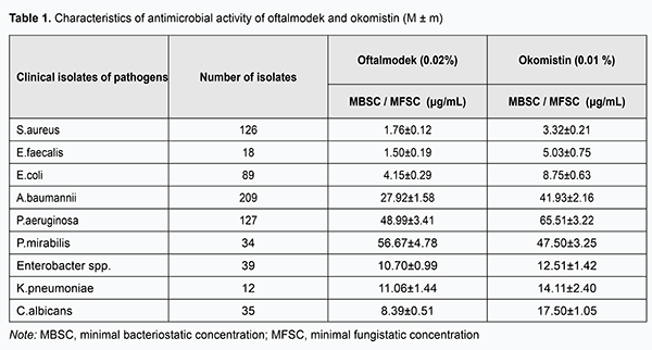
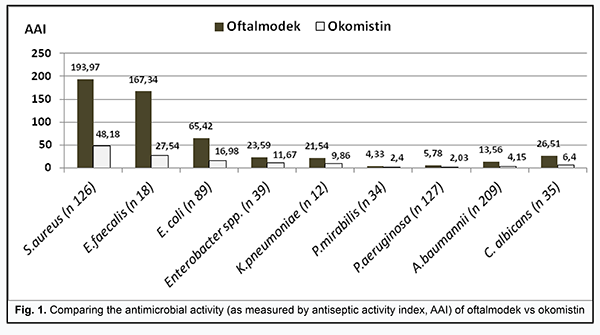
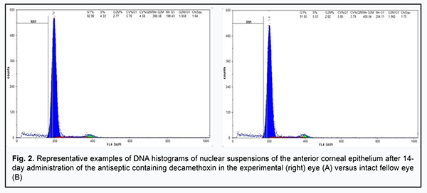
Introduction The era of antibiotic therapy and antibiotic prophylaxis began in 1929 with Alexander Fleming's report on the antibacterial effects of the Penicillium mold. Antiseptics, however, have been widely used in medical practice since as early as the mid-nineteenth century for prophylaxis and treatment of infections located at various anatomical sites, surgical scrubbing, and disinfection of instruments and rooms. The rapid growth of antibiotic industry in the twentieth century, uncontrolled administration of antibiotics, their wide use in agriculture and the capacity of microorganisms to develop resistance to antibiotics led to the healthcare crisis we have experienced over the past ten years [1]. Antiseptic medications, on the other hand, are still important in the prevention and treatment of pyogenic and inflammatory conditions and maintain their efficacy due to low pathogen resistance to these agents [2-4]. Although antibiotics have higher antimicrobial activity than antiseptics, the latter have a high antimicrobial efficacy at doses non-toxic for human tissues. As the skin and mucous membranes are more resistant against damage from chemical substances and/or medications than the tissues of the internal organs, antiseptics can be used at concentrations higher than the antibiotic dose concentrations that are safe for cells of the internal organs. Detergents (or surface-active materials) are important antiseptics [4]. The quaternary ammonium compound (QAC) antiseptics which have broad-spectrum antibacterial, antiviral and antifungal activity and are effective against resistant infectious agents are widely used in ophthalmological practice. These antiseptics have high biocompatibility indices which makes them promising for a broad range of applications [5]. Given the need for improving prevention and treatment tactics for infections, in particular, in ophthalmological practice, of particular relevance will be the comprehensive investigation of the biological safety of QAC antiseptic medications having high bacteriocidal capacity. In order to enhance our understanding of properties of the QAC antiseptic medications on the market, it is important not only to investigate antimicrobial properties of these medications, but also (a) to conduct molecular studies on their effects on epithelial DNA fragmentation and cell cycle and (b) to exclude toxic effects of their therapeutic concentrations. As flow cytometry can provide information on the molecular changes at the level of the DNA of the cell (i.e., identify the DNA content of the cell nucleus and cell cycle phase distribution), it is reasonable to use this technique to assess the cytotoxicity of medications [6]. The purpose of the study was to investigate antimicrobial efficacy of the current antiseptic medications used in the ophthalmological practice, quaternary ammonium compounds (QACs) of decamethoxin (oftalmodek or OD) and miramistin (okomistin or OM), and to assess their effects on epithelial DNA fragmentation and cell cycle in rats. Materials and Methods We conducted a microbiological study to compare the two QAC antiseptic medications produced by Ukrainian manufacturers, oftalmodek (containing 0.2 mg/mL decamethoxin; Authorization Certificate №UA/10150/01/01 issued on June 9, 2017; Order №627 of June 9, 2017) and okomistin (containing 0.1 mg/mL miramistin; Authorization Certificate №UA/7537/01/01 issued on March 5, 2014; Order №159 of March 5, 2014), for the antimicrobial efficacy and cell cycle effects. The antimicrobial efficacies of oftalmodek and okomistin were examined using serial two-fold dilutions to determine minimal bacteriostatic concentrations (MBSC) of the drugs for clinical isolates of the following opportunistic pathogens that caused inflammatory and infectious processes in patients of Eye Microsurgery Department and patients with a severe surgical pathology treated at the Regional Clinical hospital of Vinnytsia: S. aureus (n=126), E. faecalis (n=18), E. coli (n=89), Enterobacter spp. (n=39), K. pneumoniae (n=12), A. baumannii (n=209), P. aeruginosa (n=127), P. mirabilis (n=34), and C. albicans (n=35) [7]. Analysis of the antimicrobial efficacies of the finished dosage forms of the drugs was conducted through the comparison of their antiseptic activity indices (AAI) which were determined by the standardized methodology (the ratio of the concentration of active ingredient in the finished dosage form of the antiseptic to the MBSC for the relevant infection agent). The effects of oftalmodek and okomistin on epithelial DNA fragmentation and cell cycle were examined experimentally using flow cytometry. Twenty adult male Wistar rats (age, 3 months; weight, 150±15 g) were used in experiments. The animals were maintained in a dark-light cycle (12 h light: 12 h dark: lights off at 19:00) under standardized conditions at the vivarium of the National Medical University. The in vivo cytotoxic effects and the influence of oftalmodek and okomistin on the cell cycle of rat’s anterior corneal epithelial (ACE) cells were investigated. The use of the ACE for examination of the effects of medications (in particular, antiseptics) on cell cycle phase distribution and DNA fragmentation index can be explained by its natural capacity for a relatively quick cell renovation, which in turn allows us to assess changes in proliferative cell activity and apoptotic rate [8]. Rats were divided into the oftalmodek (OD) group (10 animals) and okomistin (OM) group (10 animals). The experimental eye (right eye) received a drop of OD or OM four times daily for 14 days. In all the cases, the fellow eye was intact. At day 14, after intraperitoneal anesthesia with propofol (10 mg/kg), microsurgical technique and instruments were used to harvest the entire ACE for cytometric analysis. Nuclear suspension samples from each eye were prepared for flow cytometry analyses using with CyStain DNA 2 step kit (Partec GmbH, Münster, Germany). Each nuclear suspension sample was used for documentation of 20,000 events. Flow cytometry DNA measurements were determined after DAPI staining with a Partec Pas flow cytometer using trout erythrocytes as external control cells. There were two peaks in each flow cytometric DNA histograms of nuclear suspensions of the ACE. The G1 peak was set in channel 200 of 1024 (i.e. on a linear scale which is the most suitable for the analysis of DNA content) by adjusting the gain in the relevant fluorescence channel and scale [9]. The analysis was carried out with the Partec PAS flow cytometer, and results were processed with the Partec software package (FloMax). Fractions of cells in the S phase (the part of the cell cycle where synthesis of DNA occurs), G0G1 (these components of the cell cycle have 2n DNA) phase and G2+M (tetraploid; 4n DNA is present) phase were measured. Analysis of the proliferative index (S+G2+M) and the SUB-G0G1 interval preceding the G0G1 peak (DNA fragmentation) allowed us to assess both the proliferative activity of the cells under study and the features of their apoptotic death induced by the QAC antiseptics [7-9]. Non-parametric Mann-Whitney test was used for statistical analysis of flow cytometry derived cell cycle indices of the ACE. All parameters were analyzed for right and left eyes separately. Results Oftalmodek and okomistin showed high bacteriostatic effects against a broad spectrum of opportunistic pathogens (Table 1).
The analysis of their antimicrobial activities based on antiseptic activity indices demonstrated a substantial advantage for use of the finished dosage form containing decamethoxin over that containing miramistin. Oftalmodek indices of antimicrobial activity against E. faecalis, S. aureus, E. coli, A. baumannii, K. рneumoniae and other Enterobacter spp were approximately 6-, 4-, 4-, 3.3-, 2.2-, and 2-fold higher than those of okomistin (р < 0.001 for each comparison; Fig. 1). In addition, its index of antifungal activity against C.albicans was four-fold higher than that of okomistin (р < 0.001).
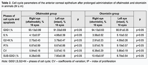
Although the resistance of P. mirabilis and P. Aeruginosa, gram-negative organisms, to oftalmodek and okomistin, was notable, the activity of the former antiseptic against these organisms was sufficient, and its respective indices of antimicrobial activity not only exceeded the minimal threshold value of 4, but were also 1.8- and 2.8-fold, respectively higher than those of okomistin (р < 0.001; Fig. 1). After having a drop of OD four times daily for 14 days, experimental eyes in the OD group demonstrated fractions of ACE cells in the S phase (the phase of DNA synthesis), and G0G1 phase (mitosis phase) that were not significantly different from those in the fellow eyes used as controls (p > 0.05, Table 2, Fig. 2).
In addition, the fraction of ACE cells in the G2+M phase in the experimental eyes in the OD group was 1.04% lower than that in the fellow eyes used as controls, but the difference was not statistically significant. Moreover, flow cytometry histogram analysis showed that the coefficients of variation (CV) were acceptable, with a CV of the G0G1 peak for the ACE in the experimental eyes in the OD group and the fellow eyes used as controls of 5.94±0.16% and 5.87±0.15%, respectively, with no significant difference (р > 0.05). Furthermore, the proliferative index of the ACE in experimental eyes in the OD group was 6.87±0.99%, which was 1.3-fold lower than that in the untreated fellow eyes. The apoptosis index (AI) of the ACE in experimental eyes in the OD group was insubstantially (by 0.68%) higher than that in the fellow eyes used as controls. The difference, however, was not significant (р > 0.05), demonstrating that a prolonged administration of OD had no proapoptotic effect on the corneal epithelium. There were differences in cell cycle phase distribution and DNA fragmentation of ACE cells between experimental eyes in the OM group and the fellow eyes used as controls. Experimental eyes in the OM group demonstrated fractions of ACE cells in the G0G1 phase that were 3.32% higher than in the fellow eyes used as controls and 2.32% higher than in experimental eyes in the OD group, and these differences were statistically significant. In addition, the CV were acceptable, with a CV of the G0G1 peak for the ACE in the experimental eyes in the OM group and the fellow eyes used as controls of 6.36±0.29% and 5.86±0.19%, respectively. Experimental eyes in the OM group demonstrated depressed DNA synthesis in nuclei of ACE cells, with fractions of ACE cells in the S phase (3.56±0.52%) that were 1.4-fold lower than in the fellow eyes used as controls (Table 2). Moreover, in experimental eyes treated with OM for 14 days, (a) the fraction of ACE cells in the G2+M phase was 1.4-fold lower than in the untreated fellow eyes (p < 0.05), and (b) the fraction of ACE cells in the SUB-G0G1 interval was 10.45±0.97% , with a statistically significant (by 3.66%) increase in DNA fragmentation level in ACE cell nuclei. Furthermore, the proliferative index of the ACE in experimental eyes in the OM group was 5.87±0.76%, which was statistically significantly lower than that (9.19±0.57%) in the fellow eyes used as controls (Table 2). Discussion Antiseptics of the QAC class contain a central nitrogen atom bound to four dissimilar alkyl groups which occur in variety of structures. They have low surface tension giving them a good ability to penetrate the microbial cell membrane. In addition, the mechanism of their antimicrobial action involves transmembrane transport of molecules and osmotic balance ions, nitrogen and phosphorus metabolism, activation of proteolytic enzymes, lysis and autolysis of organisms [10]. The current study compared the two antiseptics containing decamethoxin and miramistin, respectively, for their antiseptic activity indices for 689 clinical isolates of the opportunistic pathogens which caused infectious complications in patients of the Eye Microsurgery Department and patients with a severe surgical pathology, and demonstrated a high sensitivity of staphylococci and enterococci to these antiseptics. In addition, these antiseptics were found to have high activity indices for gram-negative enterobacteria (E. coli, Enterobacter spp., K. рneumoniae) and nonenzymatic gram-negative organisms (A. baumannii), and oftalmodek activity indices were two to four times higher than those of okomistin (р < 0.001). Although minimal inhibitory concentrations of oftalmodek and okomistin for clinical isolates of P. mirabilis and P. aeruginosa are rather high, the relevant oftalmodek activity indices were higher than the thresholds required for inhibitory effects, and 1.8 to 2.8 times higher than okomistin activity indices (р < 0.001). Current guidelines require that an antiseptic must undergo a comprehensive research process involving microbiological, biochemical, pharmacological and pharmaceutical studies. Moreover, special attention is given to toxicity studies. It is expedient to perform biological compatibility testing of an antiseptic while taking into account the results of cell cycle analysis by flow cytometry. Some surface-active antiseptics (octenisept, polyhexamethylen biguanide, chlorhexidine digluconade), although have high biological safety, have been found to be cytotoxic to epithelial cells and to inhibit proliferation of fibroblasts. Previous studies have demonstrated that chlorhexidine can stimulate stimulate apoptosis and autophagic/necrotic cell death [11]. In addition, cytotoxic effects of therapeutic concentrations of antiseptics cetylpyridinium chloride and miramistin on L929 fibroblasts and HaCaT cell lines have been demonstrated in vitro. It is still important to improve our knowledge of in vivo cytotoxic effects of surface-active antiseptics on human cells. Our experimental study found no significant difference (p > 0.05) in fractions of ACE cells in various cell cycle phases and ACE DNA fragmentation between experimental eyes that received OD and intact fellow eyes, thus evidencing no proapoptotic, antiproliferative, or cytotoxic effects of decamethoxin. Experimental eyes in the okomistin group demonstrated depressed DNA synthesis in nuclei of ACE cells, with the fraction of ACE cells in the S phase that was 1.4-fold lower than in intact fellow eyes. In addition, the fraction of ACE cells in the G2+M phase and proliferative index of the ACE were 1.4-fold and 1.6-fold, respectively, lower (р < 0.05). Moreover, flow cytometry analysis of ACE cells showed that upon prolonged administration of OD, the DNA fragmentation level in epithelial cell nuclei was increased 1.5-fold (p<0.05), providing evidence of the proapoptotic effect of the antiseptic containing miramistin. Conclusion First, oftalmodek has high antimicrobial activity against a broad spectrum of infectious agents (S. aureus, E. faecalis, E. coli, Enterobacter spp., K. рneumoniae, A. baumannii, P. aeruginosa, P. mirabilis). This was evidenced by the respective indices of its activity, which were 1.8 to 6-fold higher than those of okomistin (р < 0.001). Second, the use of oftalmodek for 14 days did not affect the corneal epithelial cell cycle parameters determined by flow cytometry. However, upon 14-day treatment with okomistin, the fraction of ACE cells capable of synthesizing nuclear DNA during the S-phase was decreased 1.5-fold, and the DNA fragmentation level in epithelial cell nuclei was increased 1.5-fold (p<0.05). Finally, prolonged administration of oftalmodek is characterized by synchronized proliferation and apoptotic death of epithelial cells, which confirms that the antiseptic has no cytotoxic or proapoptotic effects. References 1.Kamaruzzaman NF, Tan LP, MatYazid KA, et al. Targeting the Bacterial Protective Armour; Challenges and Novel Strategies in the Treatment of Microbial Biofilm. Materials (Basel). 2018 Sep 13;11(9). pii: E1705. doi: 10.3390/ma11091705. 2.Kramer A, Dissemond J, Kim S, et al. Consensus on wound antisepsis: update 2018. Skin Pharmacol Physiol. 2018;31(1):28–58. 3.Nazarchuk OA. [Modern strategy of struggle with causing agents of the infection complications]. Klin Khir. 2016;(9):59-61. Available from: https://www.ncbi.nlm.nih.gov/pubmed/30265488 4.Nazarchuk AA, Vernygorodskyi SV, Palii VG, Nazarchuk GG. [Experimental research of effectiveness of antimicrobial surgical materials containing decamethoxinum]. Novosti khirurgii. 2018;26(1):16-23. 5.Kanazawa, A., Ikeda T., Endo T. A novel approach to mode of action of cationic biocides: morphological effect on antibacterial activity. J Appl Bacteriol. 1995 Jan;78(1):55–60. 6.Voitkova VV. [Study of apoptosis with the use of flow cytometry (review of literature)]. Bulletin of the East Siberian Scientific Center SB RAMS. 2010; 76(6 Part 1): 220-225. Russian. 7.Leclercq R, Cantón R, Brown DF, et al. EUCAST expert rules in antimicrobial susceptibility testing. Clin Microbiol Infect. 2013 Feb;19(2):141-60. 8.Haddad A. Renewal of the rabbit corneal epithelium as investigated by autoradiography after intravitreal injection of 3H-thymidine. Cornea. 2000 May;19(3):378–83. 9.Brotherick I. Basic DNA Measurement by Flow Cytometry. In: Guide to flow cytometry. Wulff S, editor. Dako: Carpinteria. p. 99–106. 10.Mashkovskii MD. [Pharmaceuticals]. 15th ed, revised. Moscow: Novaia volna; 2007. Russian. 11.Karpiński TM, Szkaradkiewicz AK. Chlorhexidine – pharmaco-biological activity and application. Eur Rev Med Pharmacol Sci. 2015;19(7):1321–6. Available from: https://www.ncbi.nlm.nih.gov/pubmed/25912596 The authors certify that they have no conflicts of interest in the subject matter or materials discussed in this manuscript.
|