J.ophthalmol.(Ukraine).2017;5:12-14.
|
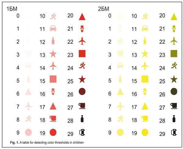
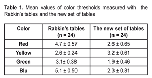
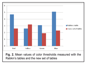
https://doi.org/10.31288/oftalmolzh201751214 A new set of tables for detecting thresholds of color sensitivity in children with acquired myopia A.Yu. Mukhina1, I.M. Boichuk2, L.D. Zhuravleva1 1Regional children clinical hospital, Ivano-Frankovsk, Ukraine 2SI “Filatov Institute of Eye Diseases and Tissue Therapy of the NAMS of Ukraine”, Odessa, Ukraine E-mail: iryna.ods@gmail.com Background. Color vision, like visual acuity, is a function of cone cells in the retina of the eye. Development and improvement of this function goes in parallel with central vision development. The main cause of impaired vision in children with acquired myopia, aside from high myopic refraction, is incomplete, in a certain degree, development of the visual system. It is difficult to diagnose this pathology. Detection of color thresholds is important for assessing the degree of incomplete development of the visual system as well as for differentiating it from congenital color vision defects. The purpose. To objectify the method and to shorten the time required for testing the thresholds of color vision using a proposed new set of tables. Material and Methods. Thirty-five patients, aged 5 to 12 years old, with moderate and high myopia of both eyes which averaged (5.6±SD2.0) D. No changes were seen in the fundus of the eye. All children were performed ophthalmic examination including visual acuity test, refractometry, and binocular function test. Color threshold were detected using the proposed set of tables (a utility model No 114858 from March, 27, 2017; Bul. No 6) and using the commonly-used Rabkin’s tables. Results. We received the mean values of threshold as follows: 4.7±0.57 c.u. to red color, 2.6±0.24 c.u. to yellow, 3.1±0.38 c.u. to green, and 5.1±0.50 c.u. to blue color using the Rabkin’s tables; 2.6±0.65 c.u. to red color: 3.2± 0.61 c.u. to yellow, 1.9± 0.46 c.u. to green, and 2.3±0.81 c.u. to blue color using the proposed tables. Conclusions. The comparison of the two methods showed that the proposed set of tables for testing thresholds of color vision simplify the test performance and minimize the time required for its passing. Also, it gives more objective and more accurate assessment of color perception and can be used for young children. Key-words: thresholds of color vision, acquired myopia, amblyopia with myopic refraction, color vision threshold test in children. Background Color vision, like visual acuity, is a function of cone cells in the retina of the eye. Development and improvement of this function goes in parallel with central vision development. The main cause of impaired vision in children with acquired myopia, aside from high myopic refraction, is incomplete, in a certain degree, development of the visual system [2]. It is difficult to diagnose this pathology. Detection of color thresholds is important for assessing the degree of incomplete development of the visual system as well as for differentiating it from congenital color vision defects. There are a number of devices and methods designed for color vision testing, most of which are focused on the color perception assessment; they include, for instance, various modes of the L?scher test, color plates designed by Schaaf, Stilling, Ishihara, Rabkin, Fletcher, Yustova and Alekseeva, colorimetry, chromography, J. Mollon test, color campimetry etc. [3, 4, 6, 7, 9, 10]. Color campimetry is a specific and highly sensitive method; however, it has some disadvantages limiting the sphere of its application and the diagnostic value. The duration of one test is about 30-40 minutes which is unacceptable in outpatient reception conditions. This method is designed for well-educated staff and requires special training by highly-skilled specialists; besides, the equipment is expensive [5]. There are two main methods which are used for color vision testing in children: special pigment tables and spectral devices, anomaloscopes. Of the pigment tables, the most perfect are recognized the polychromatic tables designed by Rabkin. The tables make it possible to detect not only a type but a degree of color perception defects. The table design is based on the principle of equation of brightness and saturation. Each table consists of dots of main and additional colors. The main color dots make up a figure or a symbol against the background of the additional color dots. The brightness of the dots is the same. Some tables have invisible figures and symbols which can be detected if vision perception disorders occur and are not seen when color vision is normal [7]. It is known that the Rabkin’s polychromatic tables are rather complicated for detecting color thresholds in children and are based on the patient’s answers, in other words, this method is subjective since children do not always understand the procedure of the test, which requires the time. This necessitates the search for new methods for assessing the thresholds of color vision in children. The purpose of the present paper was to objectify the method and to shorten the time required for testing the thresholds of color vision in young children using a proposed new method of color threshold detection. Material and Methods All children were performed ophthalmic examination including visual acuity test, refractometry, and binocular function test. Color threshold were detected using a proposed set of tables and commonly-used Rabkin’s tables. Thirty-five patients, aged 5 to 12 years old, with moderate and high myopia of both eyes, which averaged (5.6±SD2.0) D, were examined. No changes were seen in the fundus of the eye. To assess the color thresholds, we developed and proposed a new set of the tables and a method for assessing color thresholds in children (a utility model No 114858 from March, 27, 2017; Bul. No 6). The proposed method is performed as follows: a patient is shown test tables containing test elements in a form of recognizable things and geometrical shapes. Each table consists of 30 test elements. A zero test element is of minimal color saturation and of minimal contrast to the test table background. The last test element is of maximally sutured color and of maximal contrast. Each test element differs from the previous one by gradually increasing color saturation, beginning from hardly distinguishable to maximally saturated and maximally contrast to the test table background, with a step which is easily perceptible to the healthy eye. Changes in the intensity and contrast of each following test element are defined by changes in the amount of corresponding color components. In an RGB (red, green, blue) color model, it will look like a deduction of the corresponding color components (red, green, blue) from their full amount (white color). The peculiarity of the RGB color model is that, like in optometry, this color model has a strict assigned wave length for each of the three colors [1]. Three monochromatic wave lengths are defined for standard colors as R = 700.0 nm, G = 546.1 nm, B = 435.8 nm (CIE RGB); and it is supposed that any wave length will evoke the same subjective sensations as a combination of standard colors of various intensity [8]. Overview of the proposed test table for color threshold assessing is given in Figure 1.
The test was performed in conditions of daylight, with full correction, and at the distance of 30-40 sm. If a child can distinguish a figure against the white background, this is a minimal threshold of color saturation which can be perceived by the child’s eye [1]. Results We comparatively analyzed the color thresholds obtained using the two methods in the same children. The data on the color threshold comparative analysis showed no significant difference between groups, р>0.05. Little less values of the threshold were noted when using the new method; the findings are given in Table 1 and in Figure 2.
Conclusions
The comparison of the two methods showed that the proposed set of tables for color vision threshold testing simplify the test performance and minimize the time required for its passing. Also it gives more objective and more accurate assessment of color perception and can be used for young children. References 1. Boichuk IM, Mukhina AYu, Zhuravleva LD. [A method of detection of color threshold in young childlen, a model]. A utility model No 114858 from March, 27, 2017; Bul. No 6. (In Ukrainian). 2. Izmailov ChA. [Psychophysiology of colour vision]. M.: Izdatelstvo MGU; 1989. (In Russian). 3. Kvasova MD. [Vision and heredity]. M.-SPb.: Izdatelstvo Dilya Publishing; 2002. (In Russian). 4. Milanich AI. [A method of numerical detection of color vision and a device for its realization]. Patent of RF No 2499543 from 04.02.2011. (In Russian). 5. Nesteriuk LI, Vladimirova SV. [A method of detection of changes in visual system color perception. Patent of RF No 2212183 from 20.09.2003. (In Russian). 6. Padham Ch., Sonders G. [Light and color perception]. M.: Mir; 1978. (In Russian). 7. Rabkin E. B. [Polychromatic tables for color perception test]. 2005. 48p. (In Russian). 8. Rusinov MM, Grammatin AL, Ivanov PD et al. [Optical computing. Reference book]. M.: Izdatelstvo LKI; 2008. 98 p. (In Russian). 9. Shcherbakov VI, Lekomtseva AA, Polianskii AE, Parenko AE, Egorova IuV. [A method of human color vision testing]. Patent of RF No 2360592 from 10.07.2009. (In Russian). 10. Shcherbakov VI, Lekomtseva AA, Shtyrlin DA, Parenko MK, Alymov VA, Egorova IuV. [A method of human color vision testing]. Patent of RF No 2427312 from 27.08.2011. (In Russian).
|