J.ophthalmol.(Ukraine).2017;5:3-7.
|
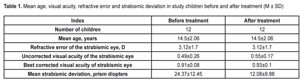
https://doi.org/10.31288/oftalmolzh2017537 Changes in the neuromuscular system of the lateral extraocular muscles after electrical stimulation of the lateral rectus muscle in children with nonaccommodative comitant esotropia V.P. Mazur, MD, I.M. Boichuk, Dr Sc (Med) Filatov Institute of Eye Disease and Tissue Therapy Odessa, Ukraine E-mail: iryna.ods@gmail.com Background: Electrical stimulation of the lateral rectus muscles (LRMs) is a physiotherapeutic technique used in the treatment of comitant esotropia. Post-treatment changes in the neuromuscular system of the eye have been not estimated previously due to the lack of an appropriate method for investigating the electrical activity of the extraocular muscles (superficial electromyography (sEMG) technique). Purpose: To employ the recently developed ocular sEMG technique in order to assess changes in the neuromuscular system of the eye in comitant esotropia before and after electrical stimulation of the LRMs. Materials and Methods: Twelve children (24 eyes) with comitant esotropia underwent electrical stimulation of the LRMs using the Amplipuls-5 apparatus. In addition, an electromyography recorder (M-TEST-2) was used to assess the function of the muscles in accordance with our sEMG methodology before and after treatment. Results: After treatment, the amplitude of the sEMG signal from the LRM insignificantly decreased from 11.55 ±2.3 to 10.64 ± 0.7 mV (p > 0.05), whereas the frequency significantly increased from 49.7 ± 3.6 to 63.18 ± 8.2 Hz (p < 0.05). In addition, the amplitude and frequency of the sEMG signal from the medial rectus muscle insignificantly (p > 0.05) decreased, from 11.48 ± 0.5 mV to 10.2 ± 0.9 mV and from 101.96 ± 5.6 Hz to 94.7 ± 19.5 Hz, respectively. Conclusion: The sEMG technique allows for estimating changes in frequency and amplitude of the response of the LRMs before and after electrical stimulation. In children with comitant esotropia, an improvement in the imbalance between the activity indices of lateral and medial rectus muscles was observed after electrical stimulation of the extraocular muscles. Keywords: electrical stimulation of extraocular muscles, superficial electromyography, comitant esotropia Introduction Most ophthalmologists consider only orthoptic and pleoptic treatment, but not physiotherapeutic methods when managing patients with comitant esotropia. However, the potential of electrical stimulation therapy for patients with different types of strabismus is rather high. Low awareness of physiotherapeutic approaches for the treatment of strabismus among practitioners may be explained not only by unavailability of proper literature, but also by their poor knowledge of the mechanisms of action of electric pulses on injured muscles and of the ways to control the efficacy of treatment. Yurov [1] and Cherikchi and colleagues [2] from the Department of Physiotherapy at the Filatov Institute published recommendations on the treatment of comitant esotropia with electric pulses in 1968 and in 1964, respectively. They had reviewed the works of Fisher (1958) [1] and Pilman (1959) [2] on the importance of primary injury to extraocular muscles (in the form of paresis of the lateral rectus muscles (LRMs)) in the mechanism of the development of comitant esotropia. In addition, they found this importance had been confirmed in the study by Fridman (1964) [1], with the paretic component revealed in 74% of the comitant esotropia cases. This is a potential reason for the absence of effect with common orthoptic methods in most patients, and, therefore, justifies the use of physiotherapeutic approaches in comitant esotropia cases [1, 3-5]. The features of the neuromuscular system of the eye impose limitations on electric diagnostic studies, because the current required for a visible deflection of the eye is substantially higher than the human pain threshold. Therefore, Cherikchi and colleagues could judge the patient’s muscle response to electrical stimulation only by fibrillary muscle tremor felt by the patient. The potential of individual muscle fibers for contraction has been confirmed by the direct observation of the response of the animal muscle subjected to the recommended mode of electrical stimulation. The data obtained provided evidence that the effect of electrical stimulation of the muscles is based on “electric gymnastics” approaches [2]. The magnitude of deviation of the strabismic eye was not the only aspect used by those authors to assess the therapeutic effect of electrical stimulation of extraocular muscles. Changes in the state of the neuromuscular system of the eye were assessed also through muscle coordination measurements that allow estimating the motility of the muscle of the strabismic eye. As the results of that study depended on the patient’s psychophysiological state, eye movement amplitude and speed, and on the amount of impairment of retinal function, that methodology is not sufficiently reliable, and does not allow for a comprehensive understanding of the changes in the neuromuscular system of the eye after a course of electrical stimulation. Previously, we have developed a superficial electromyography (SEMG) technique [6] that allows for comprehensive assessment of total bioelectrical activity of the LRMs of the strabismic eye before and after treatment. The purpose of this study was to employ the previously developed ocular sEMG technique in order to assess changes in the neuromuscular system of the eye in comitant esotropia before and after electrical stimulation of the LRMs. Materials and Methods Twelve children (24 eyes) with comitant esotropia underwent ocular examination and treatment. They had neither disturbances of the optic media nor retinal disorders. Data from history taking and ocular examination were used to classify the type of strabismus according to the Rykov and Synyakina classification of 2008. Table 1 shows patients’ characteristics before and after treatment.
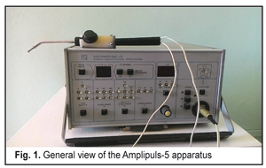
An electromyography recorder (M-TEST-2) was used to assess the function of horizontal recti in accordance with the methodology that we have reported previously [6]. Two 5-mm silver-and platinum SEMG electrodes placed at an interelectrode distance of 6 mm were used to record SEMG signals. The active electrode was placed on the muscle belly (with the eye maximally retracted) and the reference on the limbus. The study was conducted after the muscles were anesthetized with Alcain 0.5% in the following order: first, the lateral rectus muscle of the right eye, and the medial rectus of the right eye, with the eyes in maximal levoversion and dextraversion; and, second, the lateral rectus muscle of the left eye, and the medial rectus of the left eye, with the eyes in maximal dextraversion and levoversion. The following SEMG parameters were recorded in triplicate for each rectus muscle: frequency of the total electrical activity of the muscles, maximum amplitude of the SEMG signal, and mean amplitude of the SEMG signal. Data was processed automatically. The Amplipuls-5 apparatus (MAYAK, FGUP (100% state-owned company), Kursk, Russia) was used for electrical stimulation of the horizontal rectus muscles (Fig. 1).
Methodology Three drops of anesthetic agent, Alcain 0.5%, were administered with 1-min intervals. Thereafter, electrical stimulation of the lateral rectus muscle (LRM) and of the medial rectus muscle (MRM) was performed in comitant esotropia and exotropia, respectively, with the electrode placed 5-6 mm from the limbus. Table 2 shows the therapeutic regimen proposed by Yurov and Cherikchi.
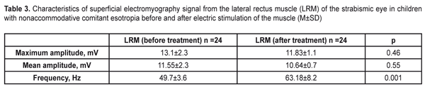
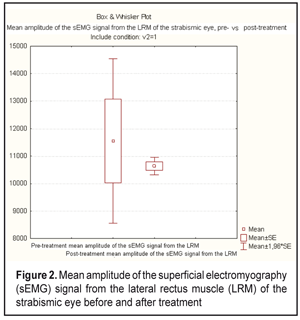
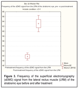
Results and Discussion The data on the electrical activity of the LRMs of the strabismic eye before and after treatment is presented in Table 3 and Figs. 2 and 3. The table shows that the amplitude of SEMG signal from the LRMs decreased (p > 0.05), whereas the frequency of SEMG signal significantly increased (p <0.05) after treatment.
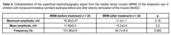
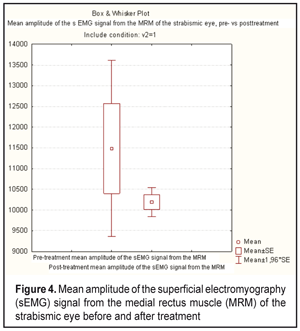
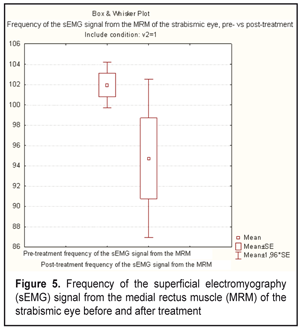
The data on the electrical activity of the medial rectus muscles of the strabismic eye before and after treatment is presented in Table 4 and Figs. 4 and 5. The table shows that the frequency and the amplitude of SEMG signal from the medial rectus muscles decreased (p > 0.05) and became more normal after treatment.
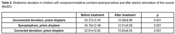
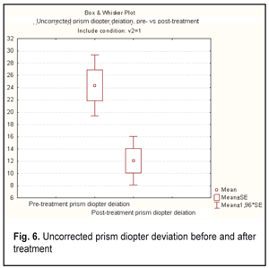
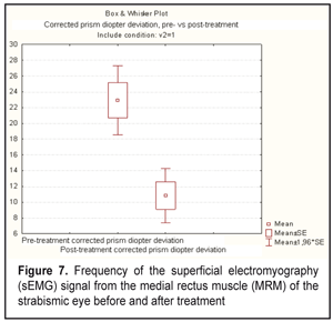
The data on the magnitude of strabismic deviation in study patients before and after treatment is presented in Table 5 and Figs. 6 and 7. The table shows that the magnitude of strabismic deviation in study patients significantly improved (p < 0.05) after treatment.
Conclusions First, the superficial electromyography technique allows for estimating changes in frequency and amplitude of the response of the LRMs of the eye before and after treatment. Second, in esotropia, the frequency of the sEMG signal from the LRM increased from 49.7±3.6 Hz to 63.18 ± 8.2 Hz (p < 0.05) after electrical stimulation of the muscle. Third, in esotropia, the total amplitude of the sEMG signal from the LRM decreased from 13.1 ± 2.3 mV to 11.83 ± 1.1 mV (p > 0.05) after electrical stimulation of the muscle. Fourth, in esotropia, activity indices (frequency and amplitude of the sEMG signal) of the medial rectus muscle insignificantly decreased (p < 0.05) after electrical stimulation of the LRM.
Finally, in children with comitant esotropia, an improvement in the imbalance between the activity indices of the lateral and medial rectus muscles, with improvement in the magnitude of strabismic deviation, was observed after electrical stimulation.
References 1. Yurov SI. [Treatment of comitant esotropia with electrical stimulation of the lateral rectus muscles]. Oftalmol. Zh. 1968;8: 597-601. Russian 2. Cherikchi LE. [Physical therapy in ophthalmology]. Kyiv: Zdorov’ia; 1979. Russian 3. Andriienko AA. [Electrical stimulation of extraocular muscles as a component in the comprehensive management of strabismus]. Vrach Delo. 1975;11:103-5. Russian 4. Sosin IN, Buiavykh AG. [Physical therapy for ocular disorders: a guide for practitioners]. Simferopol: Tavriia; 1998. Russian 5. Sasaki T, Suzuki K, Matsumoto M, et al. Origins of surface potentials evoked by electrical stimulation of oculomotor nerves: are they related to electrooculographic or electromyographic events? J Neurosurgery. 2002 Oct;97(4):941-4 6. Boichuk IM, Mazur VP. [A new method of surface electromyography of direct eye muscles in children]. Oftalmol. Zh. 2014;3:15-18. Russian
|