J.ophthalmol.(Ukraine).2017;4:31-39.
|
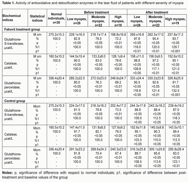
https://doi.org/10.31288/oftalmolzh201743139 Antioxidative status as assessed by enzymatic activity in the tear fluid and by the levels of sulfur-containing protein groups in the blood and tear fluid of myopes before and after treatment with a thiol agent E.I. Surova, a Postgraduate student, I.M. Boichuk, Dr. of Sc. (Med.), S.G. Kolomiichuk, a research fellow Filatov Institute of Eye Diseases and Tissue Therapy; Odessa (Ukraine) E-mail: iryna.ods@gmail.com Background: Myopia remains a leading cause of vision loss and visual disability in Ukraine (75%) and one of the most common ocular disorders worldwide. Most of available biological materials like blood and tear fluid can be used to measure the levels of these compounds, and thus to assess the relative balance between lipid peroxidation (LPO) and antioxidant system (AOS) in myopes. This will enable to find a pharmacological way for normalizing this balance in order to prevent degenerative retinal changes, and to stop the progression of myopia. Purpose: To evaluate the antioxidant status in patients with different severity of myopia based on the activity of enzymes (glutathione-S-transferase (GST), catalase (CAT), and glutathione peroxidase (GPx)) in their tear fluid and of sulfur-containing protein groups in their blood and tear fluid before and after treatment with Fakovit, a thiol agent. Materials and Methods: The Facovit treatment group included 67 myopes (low myopes, n = 26; medium myopes, n = 22; high myopes, n = 19) aged 12-24 years, whereas the control group included 67 aged-matched who did not receive Facovit treatment. In addition, 30 aged-matched healthy individuals were included into the study. Results: At baseline, GPx neutralized lipid hydroperoxides too slowly, and GST activity was markedly inhibited, with GST activity in the tear fluid and in the blood in low, moderate and high myopes of the Fakovit treatment group being statistically significantly lower than in normal individuals. In addition, CAT activity in the tear fluid in moderate and high myopes of this group was low. The baseline levels of free and protein-bound sulphydryl and disulphide groups in the tear fluid in low, moderate and high myopes of the Fakovit treatment group were abnormal, indicating substantial abnormalities of the AOS, with the tear fluid levels of free sulfhydryl groups and of protein-bound sulfhydryl groups being statistically significantly lower, and with the tear fluid levels of free disulphide groups and of protein-bound disulphide groups being higher than in normal individuals. Biochemical study demonstrated that the treatment including the thiol agent resulted in a substantial increase in the activity of enzymes (GPx and GST) of the detoxification system. Consclusion: Fakovit was found to have a substantial antioxidation effect on thiols in myopia, with increase in the levels of free sulfhydryl groups and decrease in the levels of protein-bound disulphide groups in the blood and tear fluid. Key-words: myopia, antioxidation system, enzymatic activity in the tear, level of sulfur-containing protein groups in blood and tear fluid, Fakovit, a tiol agent Introduction Myopia remains a leading cause of vision loss and visual disability in Ukraine (75%) and one of the most common ocular disorders worldwide. Clinical studies have demonstrated that more than 40% of children and adolescents with progressive myopia develop central or peripheral chorioretinal dystrophy, with the incidence of this dystrophy substantially increasing with age, severity and duration of myopia. Abnormal metabolism in the scleral connective tissue, choroid and retina results in further progression of myopia with the development of irreversible desctructive changes followed by vision loss, and, subsequently, by limitations in choice of profession and by disability [1]. Activation of lipid peroxidation (LPO) in the sclera and tear of patients with progressive myopia has been demonstrated in numerous studies [1-7]. Antioxidants, the substances protecting the body from free radicals, are divided into enzymatic and nonenzymatic. Free radicals can destroy biological molecules, including nucleic acids, proteins, carbohydrates and lipids, in enzymatic reactions following oxidative stress. The compounds formed as a result of these reactions can become biomarkers of the balance between LPO and antioxidant system (AOS). Most of available biological materials like blood and tear fluid can be used to measure the levels of these compounds in myopes, and thus to evaluate the relative balance between the systems. This will enable to find a pharmacological way for normalizing this balance in order to prevent degenerative retinal changes and to stop the progression of myopia. There have been reports on studies on the biochemical composition of tear fluid in various ocular disorders [1,4,6,8]. Previously we have reported on the analysis of changes in the antioxidant status based on the blood enzyme levels in myopes following treatment with Fakovit, a thiol agent [9]. It should be noted that the level of free SH groups determines the level of reduced glutathione, whereas the level of free SS groups determines the level of oxidized glutathione. Currently, the extent to which the levels of free and protein-bound SH and SS groups in the tear fluid and blood deviate from the norm is considered essential for antioxidant defense. To our best knowledge, antioxidant status has never been studied in the tear fluid of patients with different severity of myopia, and this is the first study to report the antioxidant status in the tear fluid and blood of such patients before and after treatment with a thiol agent. The purpose of this study was to evaluate the antioxidant status in patients with different severity of myopia based on the activity of enzymes (glutathione-S-transferase (GST), catalase (CAT), and glutathione peroxidase (GPx)) in their tear fluid and of sulfur-containing protein groups in their blood and tear fluid before and after treatment with Fakovit, a thiol agent. Materials and Methods The Facovit treatment group included 67 myopes (low myopes, n = 26; medium myopes, n = 22; high myopes, n = 19) aged 12-24 years, whereas the control group included 67 aged-matched myopes who did not receive Facovit treatment. In addition, 30 aged-matched healthy individuals were included into the study. Fakovit treatment regimen was as follows: a tablet of each type (those dissolved in the stomach and those dissolved in the gut) twice a day with meals within a 30-day treatment course, three to four courses a year [9]. All patients underwent ophthalmic examinations including refractometry, biomicroscopy, echobiometry, ophthalmoscopy, and absolute reserve of accommodation. The uncorrected and corrected distance visual acuities were assessed using the Shevalev’s Chart, and absolute reserve of accommodation was assessed using a conventional technique. Biochemical studies included assessment of the activity of GST, CAT, and GPx in the tear fluid, and determination of thiol (sulphydryl) and disulphide groups in the tear fluid and blood before and after treatment with Fakovit. Antioxidant status was assessed through the activity of enzymes in the tear fluid before and after treatment with Fakovit. GST is involved in the transfer of aminoacids through membanes and in the reduction of proteins, and its activity was assessed using a Specol-210 (Carl Zeiss, Jena, Germany) spectrophotometer at 360 nm. CAT is involved in the detoxification of LPO products, and its activity was assessed using an SF-26 (Lomo, St Petersburg, Russia) spectrophotometer at 230 nm. GPx is involved in the protection of lipid membranes from oxidative damage, and its activity was assessed with the method of Moin using an SF-16 (Lomo) spectrophotometer at 412 nm [10]. The levels of thiol (sulphydryl and disulphide) groups in the tear fluid and blood were determined based on the amount of thionitrophenol released through the reaction of 5,5?-dithiobis(2-nitrobenzoic acid) (Ellman's reagent) with free protein SH groups. Optical density of solutions was measured at 412 nm on a Specol-210 spectrophotometer. The levels of thiol (sulphydryl and disulphide) groups in the tear fluid or blood were expressed in ?mol/l [11]. STATISTICA 8 software package was used for statistical analysis. Results Table 1 presents data on the activity of enzymes of the antioxidant and detoxification systems in the tear fluid of patients with different severity of myopia of the Facovit treatment and control groups.
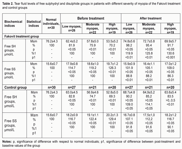
At baseline (before treatment), in the Fakovit treatment group, the activity of enzymes was lower than in normal individuals (275.2±15.3 ?kat/l), the amount of abnormality depending on the severity of myopia. Thus, the tear fluid GST activity in low, moderate and high myopes was 17.1% (p < 0.05), 21.5% (p < 0.05), and 27.8% (p < 0.01), respectively, lower than in normal individuals. The tear fluid CAT activity in moderate and high myopes was 17% (p < 0.05) and 21.3% (p < 0.05), respectively, lower, whereas the tear fluid GPx activity in low, moderate and high myopes was 20% (p < 0.05), 23.7% (p < 0.01), and 31.8% (p < 0.01), respectively, lower than in normal individuals. After treatment with Fakovit, in low, moderate and high myopes, the tear fluid GST activity increased by 18.0% (p < 0.01), 20% (p < 0.05), and 29.7% (p < 0.001), respectively; the tear fluid CAT activity increased by 9.8% (p < 0.05), 17.1% (p < 0.01), and 21.0% (p < 0.01), respectively; and the tear fluid GPx activity increased by 16.8% (p < 0.05), 21.4% (p < 0.01) and 32.5% (p < 0.001), respectively. Although the activity of enzymes in the tear fluid after treatment increased compared to baseline values, it was still lower than normal. At baseline (before treatment), in the control group (Table 1), the activity of enzymes was lower than in normal individuals (224.2±17.0 ?kat/l). Thus, in low, moderate and high myopes of the control group, the tear fluid GST activity was 19.5%, 21.4%, and 27.5%, respectively, lower; the tear fluid CAT activity was 8.3%, 17.9%, and 20.5%, respectively, lower; and the tear fluid GPx activity was 18.5%, 24.6%, and 22.6%, respectively, lower than in normal individuals. After conventional treatment (without the thiol agent), the amount of improvement in tear fluid enzyme activity depended on the severity of myopia, and, in low, moderate and high myopes of the control group, the tear fluid GST activity increased by 9%, 12.5% and 18.3% (p < 0.05), respectively; the tear fluid CAT activity increased by 3.5% ( p > 0.05), 10.0% (p < 0.05) and 12.5% (p < 0.05), respectively; and the tear fluid GPx activity increased by 8.5% (p <0.05), 13.6% (p < 0.05), and 23.1% (p < 0.01), respectively, compared to baseline values, but was still lower than normal. Therefore, after treatment, in the Fakovit treatment group (in patients with any severity of myopia), the tear fluid enzyme activities increased (18%-32.5%, p < 0.01) more and were closer to normal than those in the control group (3.5%-23%, p < 0.05). This fact evidences that treatment of our myopic patients with the thiol agent promoted normalization of the enzymatic component of the AOS in the tear fluid. Table 2 presents data on the levels of free sulphydryl and disulphide groups in the tear fluid of patients with different severity of myopia of the Fakovit treatment and control groups.
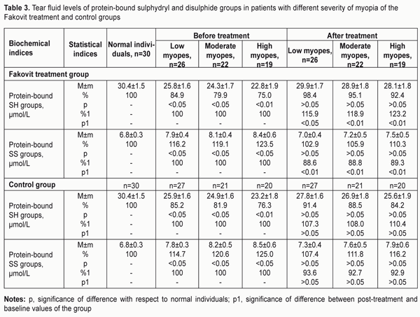
At baseline (before treatment), in low, moderate and high myopes of the Fakovit treatment group, the tear fluid levels of free sulfhydryl (SH-) groups were 19.1%, 24.1%, and 29.8%, respectively, lower than in normal individuals. After treatment, in low, moderate and high myopes of the Fakovit treatment group, the tear fluid levels of free sulfhydryl groups increased by 19.9% (p < 0.01), 25.8% (p < 0.01) and 30.6% (p < 0.001), respectively, and became normal. At baseline (before treatment), in low, moderate and high myopes of the Fakovit treatment group, the tear fluid levels of free disulphide (SS-) groups were 14.7% (p < 0.05), 19.2% (p < 0.05) and 26.3% (p < 0.01), respectively, lower than in normal individuals. After treatment, in low, moderate and high myopes of the Fakovit treatment group, the tear fluid levels of free disulphide (SS-) groups increased by 11.2%, 11.8% and 13.7%, (p < 0.01), respectively, and became normal. At baseline (before treatment), in low, moderate and high myopes of the control group, the tear fluid levels of free sulfhydryl (SH-) groups were 17.2% (p < 0.05), 25.3% (p < 0.01) and 30.7% (p < 0.01), respectively, lower than in normal individuals. After treatment, in low, moderate and high myopes of the control group, the tear fluid levels of free sulfhydryl (SH-) groups increased by 9%, 14.1% and 20.5%, (p < 0.01), respectively, and became normal. At baseline (before treatment), in low, moderate and high myopes of the control group, the tear fluid levels of free disulphide (SS-) groups were 16.7% (p < 0.05), 22.4% (p < 0.05) and 29.4% (p < 0.01), respectively, lower than in normal individuals. After treatment, in low, moderate and high myopes of the control group, the tear fluid levels of free disulphide groups increased by 8.2%, 8.4% and 9.9%, (p < 0.05), respectively, and became closer to normal. Overall, the data of Table 2 demonstrate normalization of the sulfur-containing protein component of the AOS in the tear fluid of myopic patients after treatment, with the improvement being more pronounced in patients treated with a thiol agent than in conventionally treated patients. Table 3 presents data on the levels of protein-bound sulphydryl (SH-) and disulphide (SS-) groups in the tear fluid of patients with different severity of myopia of the Fakovit treatment and control groups.
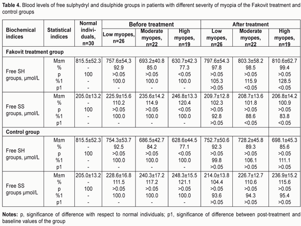
The table shows that, at baseline (before treatment), in low, moderate and high myopes of the Fakovit treatment group, the tear fluid levels of protein-bound sulphydryl groups were 15.1% (p < 0.05), 20.1% (p < 0.05) and 25% (p < 0.01), respectively, lower than in normal individuals (30.4±1.5 ?mol/L). After treatment with a thiol agent (Fakovit), in low, moderate and high myopes of this group, the tear fluid levels of protein-bound sulphydryl groups increased by 15.9% (p < 0.05), 18.9 (p < 0.01) and 23.2% (p < 0.01), respectively, compared to baseline. At baseline (before treatment), in low, moderate and high myopes of the Fakovit treatment group, the tear fluid levels of protein-bound disulphide (SS-) groups were 16.2%, 19.1% and 23.5% (p < 0.05), respectively, higher than in normal individuals (30.4±1.5 ?mol/L). After treatment with Fakovit, in low, moderate and high myopes of this group, the tear fluid levels of protein-bound disulphide groups decreased by 11.4%, 11.2 and 11.7% (p < 0.01), respectively, and became closer to normal. At baseline (before treatment), in low, moderate and high myopes of the control group, the tear fluid levels of protein-bound sulphydryl (SH-) groups were 14.8% (p < 0.05), 18.1% (p < 0.05), and 23.7% (p < 0.01), respectively, lower than in normal individuals. After conventional treatment, in low, moderate and high myopes of this group, the tear fluid levels of protein-bound sulphydryl groups decreased by 7.3%, 8.0 and 10.4%, respectively, but the decrease was not significant (р > 0.05). At baseline (before treatment), in low, moderate and high myopes of the control group, the tear fluid levels of protein-bound disulphide (SS-) groups were 14.7%, 20.6% and 25% (p < 0.05), respectively, higher than in normal individuals. After conventional treatment, in low, moderate and high myopes of this group, the tear fluid levels of protein-bound disulphide groups decreased by 6.3%, 7.3% and 7.1%, respectively, but the decrease was not significant (р > 0.05). Therefore, after treatment, the tear fluid levels of protein-bound sulphydryl (SH-) and disulphide (SS-) groups in the control group did not change, whereas those in the Fakovit treatment group became closer to normal, indicating an improvement in the antioxidant defense system in the tear fluid. We also evaluated the relative balance between LPO and AOS in the blood of patients with different severity of myopia based on the levels of sulfur-containing protein groups. Table 4 presents data on the levels of free sulphydryl (SH-) and disulphide (SS-) groups in the blood of patients with different severity of myopia of the Fakovit treatment and control groups.
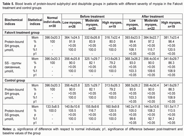
At baseline (before treatment), in the Fakovit treatment group, only high myopes demonstrated substantially (22.7%) and significantly (p < 0.05) lower levels, whereas low and moderate myopes demonstrated insubstantially (7.1% and 15%, respectively) and significantly (р > 0.05) lower blood levels of free sulphydryl groups compared to normal individuals (815.5±52.3 ?mol/L). After treatment with Fakovit, in high, moderate and low myopes, the blood levels of free sulphydryl groups increased by 28.3% (p < 0.05), increased by 15.3% (p < 0.05), and and did not change (p > 0.05), respectively, compared to baseline. At baseline (before treatment), in the Fakovit treatment group, only high myopes demonstrated substantially (20.4%) and significantly (p < 0.05) higher levels, whereas low and moderate myopes demonstrated insubstantially (10.2% and 14.9%, respectively) and insignificantly (р > 0.05) higher blood levels of free disulphide groups compared to normal individuals. After treatment with Fakovit, in low, moderate and high myopes, the blood levels of free disulphide groups decreased by 7.2% (р > 0.05), 11.4% (p < 0.05), and 16.2% (p < 0.05), respectively, compared to baseline. In addition, Table 4 demonstrates that, at baseline, in the control group, only high myopes demonstrated significantly (p < 0.05; 22.9%) lower levels, whereas low and moderate myopes demonstrated insignificantly lower levels of free sulphydryl groups in the blood compared to normal individuals. After conventional treatment of myopia, the blood levels of free sulphydryl groups did not change (р > 0.05). In low, moderate and high myopes of the control group, the blood levels of free disulphide groups were insignificantly (11.5%, 17.2% and 21.1%, respectivly; p > 0.05) higher than in normal individuals at baseline, and changed insignificantly after treatment. Overall, the data of Table 4 demonstrate that treatment of myopia with the tiol agent promotes normalization of the sulfur-containing protein component of the balance between LPO and AOS, with a decrease in the oxidized thiol levels (seen through free SH and SS group levels) in moderate anf high myopes, whereas conventional myopia treatment has no effect on these parameters. Protein-bound SH and SS groups represent thiol and disulphide bonds in protein structures, and are a key component of the antioxidant defense. Table 5 presents the blood levels of protein-bound sulphydryl and disulphide groups in patients with different severity of myopia in the Fakovit treatment and control groups.
At baseline (before treatment), in low, moderate and high myopes of the Fakovit treatment group, the blood levels of protein-bound sulphydryl groups were 8.1%, 16.1% and 20% (p < 0.05), respectively, lower than in normal individuals (396.0 ± 20.3 ?mol/L), and this difference was significant (p < 0.05) only in high myopes. After treatment with the thiol agent, in low, moderate and high myopes, the blood levels of protein-bound sulphydryl groups increased by 8.1% (p > 0.05), 15.7%, (p < 0.05) and 20.5% (p < 0.05), respectively. At baseline (before treatment), in low, moderate and high myopes of the Fakovit treatment group, the blood levels of protein-bound disulphide groups were 10%, 17.9% and 20.8%, respectively, lower than in normal individuals, and this difference was significant (p < 0.05) only in high myopes. After treatment, in low, moderate and high myopes of this group, the blood levels of protein-bound disulphide groups increased by 3.3%, 9.5% and 8.9% (p > 0.05) respectively. Table 5 demonstrates that, at baseline (before treatment), in low, moderate and high myopes of the control group, the blood levels of protein-bound sulphydryl groups were 10%, 17.9% and 21.8%, respectively, lower than in normal individuals, and this difference was significant (p < 0.05) only in high myopes. After conventional treatment, in low, moderate and high myopes of this group, the blood levels of protein-bound sulphydryl groups increased by 3.3%, 9.6% and 8.9% (p > 0.05) respectively. At baseline (before treatment), in low, moderate and high myopes of the control group, the blood levels of protein-bound disulphide groups were 5.5%, 16.7% and 20.5%, respectively, lower than in normal individuals, and this difference was significant (p < 0.05) only in high myopes. After conventional treatment, in myopia of any severity in this group, the blood levels of protein-bound disulphide groups decreased insignificantly (p > 0.05) compared to baseline values, but were still higher than normal. Overall, Table 5 demonstrates that, in myopia (especially, high myopia), AOS characteristics are reduced, but the treatment including the thiol agent allowed improving the defense capacity of the AOS, with increase in blood protein-bound disulphide levels in moderate and high myopes by 15.7% and 20.5%, respectively. Conventional treatment of myopia has no effect on these biochemical characteristics. Conclusion First, it was found that at baseline, glutathione peroxidase neutralized lipid hydroperoxides too slowly, and glutathione-S-transferase activity was markedly inhibited, with glutathione-S-transferase activity in the tear fluid in low, moderate and high myopes of the Fakovit treatment group being 17.1%, 21.5%, and 27.8%, respectively, lower than in normal individuals (the differences were statistically significant). Second, at baseline, catalase activity in the tear fluid in moderate and high myopes of this group was 17.1% and 21.3%, respectively, lower, whereas glutathione peroxidase activity in the tear fluid in low, moderate and high myopes was 20%, 23.7%, and 31.8%, respectively, lower than in normal individuals. Third, the baseline levels of free and protein-bound sulphydryl and disulphide groups in the tear fluid in low, moderate and high myopes of the Fakovit treatment group were abnormal, indicating substantial abnormalities of the AOS, with the tear fluid levels of free sulfhydryl groups and of protein-bound sulfhydryl groups being 19.1%, 24.1%, and 29.8%, respectively, lower, and 15.1%, 20.1%, and 25%, respectively, lower, and with the tear fluid levels of free disulphide groups and of protein-bound disulphide groups being 14.7%, 19.2% and 26.3%, respectively, and 16.2%, 19.1% and 23.5%, higher than in normal individuals. Fourth, our biochemical study demonstrated that the treatment including the thiol agent resulted in a substantial increase in the activity of enzymes of the detoxification system. Thus, in low, moderate and high myopes, GPx activity in the tear fluid increased by 16.8%, 21.4% and 32.5%, respectively; CAT activity in the tear fluid increased by 9.8%, 17.1%, and 21.0%, respectively; and GST activity in the tear fluid increased by 18.0%, 20%, and 20.7%, respectively. An increase in enzyme activity in the control group was observed two times less frequently than in the Fakovit treatment group. Finally, the thiol agent, Fakovit, was found to have a substantial antioxidation effect on thiols in myopia. After treatment, in low, moderate and high myopes of the Fakovit treatment group, the tear fluid levels of free sulfhydryl groups increased by 19.9%, 25.8% and 30.6%, respectively, and the blood levels of protein-bound sulphydryl groups increased by 8.1%, 15.7%, and 20.5%, respectively. In addition, the tear fluid levels of protein-bound disulphide groups decreased by 11.4%, 11.2 and 11.7%, respectively. However, in the control group, no change in the levels of sulphydryl groups and of protein-bound disulphide groups in the blood and in the tear fluid was observed after conventional treatment for myopia. References
|