J.ophthalmol.(Ukraine).2016;5:47-53.
|
https://doi.org/10.31288/oftalmolzh201654753 Effect of PFCL versus heavy silicone oil on the structure and functional state of the rabbit retina D.V. Zhmuryk,1 Cand Sc (Med) V.V. Vit,2 Dr Sc (Med), Prof. N.I. Molchaniuk,2 Cand Sc (Biol) N.I. Khramenko,2 Cand Sc (Med) M.V. Miliienko,1 MD 1 Kyiv Municipal Clinical Eye Hospital “Eye Microsurgery Center” 2 Filatov Institute of Eye Diseases and Tissue Therapy, NAMS of Ukraine Kyiv, Odessa, Ukraine Background: The use of perfluorocarbon liquids (PFCL) and heavy silicone oil (HSO) for post-vitrectomy tamponade would potentially widen the range of indications for and improve outcomes of vitreoretinal surgery. The academic literature is, however, equivocal as to the effect of these substances on the structure and functional state of the retina. Purpose: To ivestigate experimentally the effect of shor-term (14-day) tamponade with PFCL versus HSO on the structure and bioelectric functional activity of the rabbit retina over time. Materials and Methods: A total of 6 Chinchilla rabbits (12 eyes) were used in the study. All animals were subjected to ERG analysis before posterior subtotal pars plana vitrectomy. The tamponade with PFCL and with HSO was performed in the right eye and left eye, respectively, and was removed after 14 days. Retinal structures were examined with light and electron miscroscopy and the second ERG was performed at day 7, day 14 or day 30 after removal of the tamponade. Results: The analysis based on light and electron miscroscopy revealed similar effects of the two substances on the structures of the rabbit retina. Major changes were: (a) hydropic changes in the smooth endoplasmic reticulum of retinal pigment epithelium (RPE) cells, RPE mitochondria, photoreceptor inner segments, and ganglion and M?ller cells, and (b) compensation-and-restoration processes within these cells, which enable normalization of the structures. ERG recordings revealed no significant difference between the effect of PFCL and HSO on the functional activity of the rabbit retina: the amplitude of the response of the inner peripheral retina and that of the inner central retina were increased by 30.1% (supernormal ERG) and 44%, respectively, versus 43% and 64.7%, respectively. Conclusions: Short-term (14-day) tamponades with PFCL and heavy silicone oil have similar effects on on the structure and functional activity of the rabbit retina, with the changes which are reversible and non-degenerative in nature. Restoration of bioelectric activity of the retina to baseline levels after removal of 14-day PFCL and heavy silicone oil tamponades requires a longer period than 30 days. Key words: retina, ultrastructure, electroretinography, perfluorocarbon liquids, heavy silicone oil
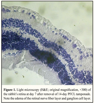
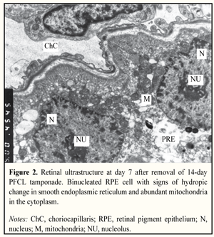
Introduction Posterior subtotal pars plana vitrectomy (PST_PPV) is still considered the gold standard for repair of retinal detachment of any cause. In PST_PPV, vitreous and fibrovascular membranes should be removed as completely as possible. In most cases, post-surgical tamponade of the vitreous cavity is required to achieve anatomical re-attachment of the retina. Light silicone oil and gas-air mixtures are effective tamponade agents in management of most retinal detachments (RDs). Surgical management of severe rhegmatogenous or traction RDs associated with inferior giant retinal tears and retinal breaks [1], anterior proliferative vitreoretinopathy (APVR) [2], or large diameter macular holes [3], however, requires heavy tamponade agents to exert the maximum tamponading effect on the inferior retina [4]. Although the use of such agents contributes to significant improvement in anatomic outcomes, the treatment does not always result in a satisfactory functional outcome, which may be associated with intraoperative trauma, toxic or mechanical damage from the tamponade agent. It is deemed important to compare the mechanical effects of perfluorocarbon liquid (PFCL) (perfluorodecalin with a specific gravity of 1.94 g/cm3) and heavier-than-water fluorinated silicone oil (specific gravity, 1.02-1.06 g/cm3) on the retina using light and electron microscopy. In addition, a highly sensitive technique (such as electroretinography (ERG)) enabling the identification of local damage to various retinal layers at pre-clinical stage may be used to assess functional alterations. The PFCL with specific gravities ranging from 1.54 g/cm3 to 1.94 g/cm3 (perfluoro-n-octane, 1.76 g/cm3; perfluortributylamin, 1.89 g/cm3; perfluorotri-n-propylamine, 1.839 g/cm3; perfluorodecalin, 1.94 g/cm3, etc.) are used in clinical practice. For an experimental study, it may be reasonable to use the PFCL with high specific gravities, since the absence of damage in the use of such compounds would mean indirectly that the use of PFCL with lower specific gravities is safe. The purpose of the study was to investigate experimentally the effect of shor-term (14-day) tamponade with PFCL versus heavy silicone oil on the structure and bioelectric functional activity of the rabbit’s retina over time. Materials and Methods A total of 6 Chinchilla adult male rabbits (12 eyes; age, 6.5 ± 0.5 months; weight, 3.5 ± 0.5 kg) were used in the study. PFCL and heavy silicone oil tamponade duration was 14 days. Each animal underwent electroretinography at the baseline (preoperatively) and at one of the three time points after removal of PFCL and heavy silicone oil tamponades. In addition, it underwent light and electron microscopy at the same time point after removal of PFCL and heavy silicone oil tamponades. After removal of the tamponades, rabbits were divided into three groups of two animals each, based on examination time point used in the study: Group 1, Group 2, and Group 3, with examination (i.e., light and electron microscopy and ERG) performed at 7, 14 and 30 days, respectively, after removal of the tamponade. Circumstances relating to animal care and experimentation met the International Guiding Principles for Biomedical Research Involving Animals as issued by the Council for the International Organizations of Medical Sciences. Operative technique Preoperatively, the rabbits were anesthetized with intramuscular thiopental sodium (2 mg/kg) and received an epibulbar injection of 0.5% proxymetacainum. Mydriasis was obtained by using one drop each of 1% atropine sulphate and 2.5% phenylephrine. Additionally, each animal received an epibulbar injection of 0.3% ofloxacinum. Posterior subtotal pars plana vitrectomy (PST_PPV) was performed under microscope observation (Zeiss OPMI 8) using a KFE-01-MEDA-NN vitrectomy system with cutting rates of 1200 cuts/min, aspiration pressure of 150 mm, and 23-G and 20-G tools. Rabbits were treated with 1.5 ml of PFCL (perfluorodecalin) and 1.0-1.5 ml of heavy silicone oil, injected into the right and left eyes, respectively. After completion of vitrectomy, 0.3% ofloxacin ointment was applied to the conjunctival sac. Removal of PFCL and silicone oil or was performed under microscope observation (Zeiss OPMI 8) using a KFE-01-MEDA-NN vitrectomy system with aspiration pressure of 150 mm Hg. Histological examination The retina was dissected from the enucleated eye, ?xed in 10% neutral-buffered formaldehyde and embedded in paraf?n. Thin sections were cut and stained with hematoxylin and eosin. Histologic evaluations of retina sections were performed by light microscopy with a Jenamed 2 2-fluorescence microscope (Carl Zeiss Jena, Germany). Inferior retinal tissue pieces were fixed in 2.5% phosphate-buffered glutaraldehyde (pH 7.4), postfixed in 1.0% phosphate-buffered osmic acid (pH 7.4), dehydrated in graded alcohols, and embedded in Epon-Araldite. Thin sections were cut, stained with Reynolds lead citrate, and examined in a PEM-100-01 electron microscope. ERG recording technique The animals were anesthetized with an epibulbar injection of 0.5% proxymetacainum. Mydriasis was obtained by using one drop each of 1% atropine sulphate and 2.5% phenylephrine. The Ganzfeld electroretinogram was performed with the Retiscan system (Roland Consult, Stasche & Finger GmbH, Brandenburg) as per ISCEV Standard for full-field clinical electroretinography comprising scotopic rod response, maximal combined scotopic response, scotopic oscillatory potentials, photopic single-flash cone response, and photopic 30-Hz flicker cone response. The full-field Ganzfeld electroretinogram (gERG) reflects the electric response of the entire retina to a flash of light (Ganzfeld stimulation is achieved with a bowl perimeter). A contact lens electrode was used for recording ERG responses. Electroretinography was performed at one of the three time points (7, 14 or 30 days) after removal of tamponade. Results Structural and functional retinal changes in two-week tamponade with PFCL At day 7 after removal of PFCL tamponade, light microscopy demonstrated edema of retinal nerve fiber layer (RNFL) and ganglion cell layer (Fig. 1), whereas electron microscopy showed no changes in the ultrastructure of the choriocapillary meshwork of uveal tissue. Fragmentation of some retinal pigment epithelium (RPE) cells and localized areas of their destruction and lysis were seen. Interestingly, that RPE cells had multiple normal organelles and some of them had two nuclei (Fig. 2). That is, signs of increased intracellular activity were observed simultaneously with evidence of destruction of mainly the membranes of smooth endoplasmic reticulum (SER). In addition, a disorder of outer segment (OS) disks and vacuolation of the mitochondria in the inner segments (IS) were seldom seen in photoreceptor cells.
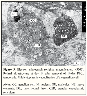
Hydropic structural changes were revealed within the internal retinal layer. Mild vacuolization of M?ller cell (MC) processes and enlargement of mitochondria occurred within the ganglion cell layer. At day 14 after removal of PFCL tamponade, most of the choriocapillaries were significantly dilated. In the microvessels, the lumen contained fine granular material of moderate electron density. RPE cells incorporated normal organelles and showed marked apical-basal polarity. Mild cytoplasmic vacuolation was observed at the expense of mitochondria and SER elements. Smooth endoplasmic reticulum membranes were swollen and exhibited localized areas of fragmentation. Within the photoreceptor layer, a portion of the cells showed evidence of edema of the inner segments and mitochondrial disorder. Photoreceptor nuclear layer and inner retinal neurons appeared normal. Marked hydropic changes along the internal retinal layer were present. Ganglion cells had nuclei with large nucleoli and contained cytoplasm with abundant granular endoplasmic reticulum (GER) and small, vacuolated mitochondria. M?ller cell processes surrounding neurons in the retinal ganglion cell (RGC) layer were characterized by mild cytoplasmic vacuolation (Fig. 3).
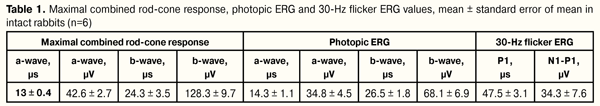
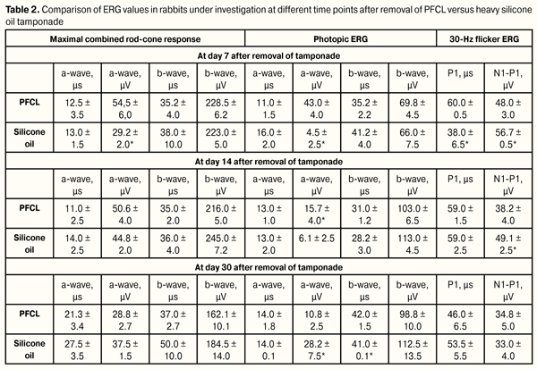
At day 30 after removal of PFCL tamponade, choriocapillaries were dilated. The lumen contents appeared normal. Although a portion of RPE cells appeared normal, other RPE cells showed pathology with cellular vacuolization and localized areas of destruction of SER elements. In addition, some RPE cells had two nuclei and abundant mitochondria with signs of activity. Localized areas of destruction of apical microvilli of RPE cells were seen. Signs of edema were present in some inner segments and damaged membrane structures were seldom seen in outer segments of photoreceptor cells. Hydropic changes were noted within the internal retinal layer. Within the ganglion cell layer, large cells had abundant organelles participating in protein synthesis, mitochondria, etc. M?ller cell processes surrounding neurons in the RGC layer showed minimal vacuolation. At day 7 following removal of PFCL, in the rabbit’s combined rod–cone response, a-wave implicit time was not significantly different from, whereas the amplitude was 28% increased (to 54,5 ± 6,0 (µV) compared to the mean baseline value, thus evidencing an increased response to the stimulus in the photoreceptor layer of the peripheral retina. In addition, the response to the stimulus in the inner retinal layers was characteristic of increased b-wave implicit time (35.2 ± 4.0 ms, p = 0.01) and amplitude (228.5 ± 6.2 µV), with the amplitude being increased by 77% compared to the mean baseline value (p = 0.001) (Tables 1, 2).
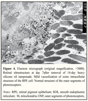
Furthermore, the photopic ERG showed no significant change in activity indices of photoreceptor layer of the central retina (with a-wave amplitude increased to 43.0 ± 4.0 µV) and inner retinal layers (with the b-wave amplitude increased to 69.8 ± 4.5 µV and the implicit time increased by 32.8%) compared to the mean baseline value (35.2 ± 2.2 ms, p = 0.001). The 30-Hz flicker ERG showed that N1-P1 amplitude of the response increased by 41% (to 48.0 ± 3.0 µV) and P1 implicit time increased by 27% (to 60.0 ± 0.5 µs) (Table 2). Therefore, a week after removal of tamponade, a response of photoreceptor and inner retinal layers of the peripheral and central retina (bipolar and M?ller cells, including those of the cone system) to the stimulation was revealed, which was reflected in increased implicit time and increased ERG amplitude (a finding of supernormal ERG). At day 14 following removal of PFCL, in the combined rod–cone response, a-wave amplitude increased by 7.4% compared to that at day 7, amounted to 54.5 ± 6.0 µV, and remained higher than that at baseline (19%, p = 0.01). In addition, the activity of the inner retinal layers (b-wave implicit time and amplitude (216.0 ± 5.0 µV) did not change significantly, and was higher than the norm (68.8%, p =0.01) (Table 2). Therefore, the activity of photoreceptor and inner retinal layers of the peripheral retina did not change significantly after day 7. Moreover, at day 14 following removal of PFCL, the photopic ERG which reflects the function of the central retina had the following features: Although the a-wave implicit time did not change, the amplitude decreased by 63.5% compared to that at day 7 and amounted to 15.7 ± 4.0 µV (p = 0.001), a factor of 2.2 lower than that the norm; the b-wave implicit time (31.0 ± 1.2 µs) (р = 0.02) remained excessive, whereas the amplitude increased by 47% compared to that at day 7, and amounted to 103.0 ± 6.50 µV (p = 0.001). The functional state of the retina at day 14 following removal of PFCL was characteristic of decreased response (depression) of central retinal photoreceptors with increased activity (irritation) of the inner layers of the whole retina. At day 30 following removal of PFCL, in the combined rod–cone response, a-wave implicit time increased from 13.0 ± 0.4 to 21.3 ± 3.4 µs (р=0.01), whereas the amplitude decreased by 34.2% compared to that at day 7, and amounted to 28.8 ± 2.70 µV (p = 0.01), thus evidencing early dystrophic changes in the peripheral retinal photoreceptors. By that day, activity values of the inner retinal layers were not restored to baseline values, and b-wave imlicit time was 30.1% higher than the norm (p = 0.01), although the amplitude was 25% lower (162.1 ± 10.1 µV) (p = 0.01) than that at day 14. In addition, no significant changes in photopic ERG measures were observed between the 14-day ERG responses and the 30-day ERG responses, although a tendency was observed towards increasing depression of the central retinal photoreceptors (Table 2) and increasing delay in response of the inner retinal layers (35% increase in b-wave implicit time; p = 0.01; the 30-day b-wave implicit time of 42.0 ± 1.5 µs), and the b-wave amplitude was elevated (98.8 ± 10.0 µV) compared to the mean baseline value (68.1 ± 6.9 µV). Therefore, at day 30 after removal of PFCL, functional activity of the central retina was characteristic of delayed response (increased implicit time) and reduced amplitude of photoreceptor response (initial dystrophy of photoreceptors) to the test flash. Inner layers of the central and peripheral retina were characteristic of increased implicit time and hyperresponsiveness to the stimulus; that is, no complete normalization of the functional state of the retina was observed and may require longer observation periods. Structural and functional retinal changes in two-week tamponade with heavy silicone oil Electron microscopy revealed that at days 7 and 14 after the removal of heavy silicone oil tamponade, the choriocapillary lumen was enlarged. In addition, vacuolation of SER was seen in RPE cells (Fig. 4). The inner and outer photoreceptor segments were swollen. Manifestations of hydropic change were seen within the outer and inner plexiform retinal layers. In addition, within the ganglion cell layer, cytoplasmic vacuolation was observed in the ganglion cells and the M?ller cells showed vacuolation of cells processes.
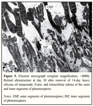
At day 30 after the removal of heavy silicone oil tamponade, fragmentation of the SER and vacuolation of mitochondria were seen in the cytoplasm of the RPE cells. The inner and outer photoreceptor segments were scattered within the photoreceptor cell layer. Disks of some outer segments were damaged. Although photoreceptors had swollen inner segments, their normal mitochondrial ultrastructure was maintained (Fig. 5). Hydropic changes were observed within the outer and inner plexiform retinal layers. Mild hydropic changes were seen in the ganglion cells. In addition, cytoplasmic vacuolation was observed in the processes of the M?ller cells located at the ganglion cells and internal limiting membrane. However, the amount of retinal nerve elements involved in retinal ultrastructural changes at day 30 after removal of heavy silicone oil was found to be substantially lower than that at day 14.
Comparative analysis of bioelectric activity of the rabbit retina after removal of a vitreous tamponade agent (PFCL or heavy silicone oil) revealed that, at day 7, in the rabbit’s combined rod–cone response, the a-wave amplitude in the silicone group was 46% lower (p = 0.001) (Table 2) than in the PFCL group, and 31.4% lower (p = 0.001) than the norm, this finding reflecting the depression in activity exhibited by the photoreceptor layer of the peripheral retina. No difference was found in bioelectric activity exhibited by the inner layers of the peripheral retina between these two groups, with this activity characterized by increased b-wave amplitude reactivity (74-77% higher than the norm). In addition, in the silicone group, there was depression in activity exhibited by the photoreceptor layer of the central retina in the photopic ERG a-wave, with the amplitude of 4.5 ± 2.5 µV being 86% lower than the norm (p = 0.001), whereas the activity of the inner layers of the central retina was normal. At day 14 after removal of tamponade, the response of the peripheral retinal photoreceptors to the stimuli in the silicone group became normal, whereas increased reactivity of the inner retinal layers in both groups maintained (Table 2). In addition, the depression in activity exhibited by the photoreceptor layer of the central retina in the silicone group remained stable (with the a-wave amplitude of 6.1 ± 2.5 µV), whereas reactivity of the inner layers of the central retina became increased (with the b-wave amplitude of 113.0 ± 4.5 µV). At day 30 after removal of heavy silicone oil, in the rabbit’s combined rod–cone response, the activity of the inner layers of the peripheral retina reduced by 24.8% (p = 0.01) compared to that at day 14, but was still higher (by 43%) compared to the mean baseline value (p = 0.01). In addition, although some positive changes were observed (the bioelectric activity of the central retinal photoreceptors improved), no changes were observed in over-reactivity of the inner retinal layers (the amplitude was 112.5 ± 4.5 µV), which was 64.7% higher than the norm (p = 0.001). Discussion A number of studies have investigated this issue. Some of them report on the development of irreversible atrophic changes in the rabbit retina following intravitreal placement of perfluoro-n-octane for 48 hours [5] or perfluorodecalin for 2 weeks [6]. One of the explanations for the difference in results between those studies and ours may be the difference in conditions for conduct of experimental studies. The use of gas compression of the vitreous by those investigators could contribute to additional damage to the retinal ultrastructure. Flores-Aguilar et al [7, 8] have reported that no morphological, functional or clinical changes in the retina and lens were observed during three months of follow-up after intraocular post-vitrectomy placement of PFCL (perflubron). According to Chang [6], after removal of 2-day perfluoro-n-octane tamponade, a-wave and b-wave amplitudes were reduced by 40% and 46%, respectively. Restoration of b-wave amplitudes to pre-vitrectomy levels began after day 5 and completed by month 2 after removal of PFCL, whereas a-wave amplitude restored to pre-vitrectomy levels by day 5 [6]. Brian et al have reported that a marked dampening of elecroretinigraphic response normalized at 14 days after removal of 24-hour or 2-week perfluorotri-n-propylamine tamponade. Therefore, our findings of the supernormal ERG response are opposed to the findings of Brian et al [9], and this is most likely due to the difference in experimental methodology. Chang et al [6] and Brian et al [9] used gas compression of the vitreous, whereas we performed PST_PPV that results in changes in the content of the vitreous cavity, with increase in concentrations of calcium, magnesium and protein and decrease in that of potassium [10]. It has been demonstrated in experimental models that the increases in a-wave and b-wave amplitudes are observed after reduction in temperature, increase in pH, and changes in ion composition in the contents of the of the vitreous cavity [11, 12]. An increase in the b-wave amplitudes of the ERG elicited by white-light or red-light stimulus at 1 month after PFCL injection into the rabbit’s vitreous has been reported by Shkvorchenko [13], who believes that these changes were associated with transudative processes. At four months after the intervention, all electroretinographic parameters restored to normal. In all the studies mentioned above, balanced salt solution was used as the control. In our experimental study, we found no significant difference between PFCL and heavy silicone oil in their effect of on the ultrastructure and functional state of the retina. Conclusions First, the features of the effects of 14-day PFCL and heavy silicone oil tamponade on the ultrastructure and functional activity of the rabbit retina were determined during the 30-day follow-up after removal of tamponade. PFCL and heavy silicone oil tamponade were found to result in similar quantitative and qualitative changes in retinal structure, which are reversible and non-degenerative in nature. Second, after removal of PFCL tamponade, the amplitude of the response of the inner peripheral retina and that of the inner central retina were increased by 30.1% and 44%, respectively. The effect of 14-day heavy silicone oil tamponade on the retina after removal of the tamponade was comparable to that of PFCL tamponade: the amplitude of the response of the inner peripheral retina and that of the inner central retina were increased by 43% and 64.7%, respectively. Restoration of bioelectric activity of the retina to baseline levels after removal of 14-day PFCL or heavy silicone oil tamponade requires a longer period than 30 days. References 1.Sirimaharaj M, Balachandran C, Chan WC, et al. Vitrectomy with short term postoperative tamponade using perfluorocarbon liquid for giant retinal tears. Br J Ophthalmol. 2005 Sep;89(9):1176-9 2.Shkvorchenko DO, Levina LV. [On the tactics of surgical treatment of proliferative diabetic retinopathy complicated by anterior proliferative vitreoretinopathy]. Oftal’mokhirurgiya. 2006;1:29-32 Russian 3.Cillino S, Cillino G, Ferraro LL, Casuccio A .Treatment of persistently open macular holes with heavy silicone oil (Densiron 68) versus C2F6. A prospective randomized study. Retina. 2016 Apr;36(4):688-94 4.Drury B, Bourke RD. Short-term intraocular tamponade with perfluorocarbon heavy liquid. Br J Ophthalmol. 2011 May;95(5):694-8 5.Devin F, Jourdan T, Saracco JB, Lucciani A. [Experimental tolerability of perfluorodecalin in prolonged intraocular tamponade]. J Fr Ophthalmol. 1995;18(4):268-74 French 6.Chang S, Sparrow JR, Iwamoto T, et al. Experimental studies of tolerance to intravitreal perfluoro-n-octane liquid. Retina. 1991;11(4): 367-74 7.Flores-Aguilar M, Munguia D, Loeb E, et al. Intraocular tolerance of perfluorooctylbromide (perflubron). Retina. 1995;15(1):3-13 8.Mackiewicz J, Maaijwee K, L?ke C, et al. Effect of gravity in long-term vitreous tamponade: in vivo investigation using perfluorocarbon liquids and semi-fluorinated alkanes. Graefes Arch Clin Exp Ophthalmol. 2007 May;245(5):665-75 9.Bryan JS, Friedman SM, Mames RN, Margo CE. Experimental vitreous replacement with perfluorotri-n-propylamine. Arch Ophthalmol. 1994 Aug;112(8):1098-102 10.Killey FP, Edelhauser HF, Aaberg TA. Intraocular fluid dynamics: measurements following vitrectomy and intraocular sulfur hexafluoride. Arch Ophthalmol. 1980 Aug;98(8):1448-52 11.Winkler BS. The electroretinogram of the isolated rat retina. Vision Res. 1972 Jun;12(6):1183-98 12.Winkler BS, Simson V, Benner J. Importance of bicarbonate in retinal function. Invest Ophthalmol Vis Sci. 1977 Aug;16(8):766-8 13.Shkvorchenko DO. [Comprehensive surgical management of retinal detachments complicated by giant retinal dialysis and tears with the use of perfluorocarbon liquids] [Cand Sc (Med) Thesis]. Moscow: Fyodorov Eye Microsurgery Complex; 1995. 132 p. Russian
|