J.ophthalmol.(Ukraine).2016;4:20-22.
|
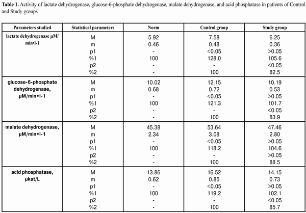
https://doi.org/10.31288/oftalmolzh201642022 Effect of sodium hyaluronate on the pathochemical process in the anterior eye segment when using benzalkonium chloride eye drops in glaucoma patients V.I. Senishin, MD Danylo Halytsky Lviv National Medical University Lviv, Ukraine E-mail: oko_science@mail.ru Introduction.The purpose of the present paper was to study the effect of sodium hyaluronate on the pathochemical process in the anterior eye segment when using benzalkonium chloride eye drops in glaucoma patients Material and Methods. The patients were divided into two groups: Control group consisted of 25 glaucoma patients being instilled Latanoprost with benzalkonium chloride (BAC); Study group consisted of 36 patients being additionally instilled 0.1% sodium hyaluronate solution for 6 months. In the tear fluid we determine the activity of lactate dehydrogenase (LD), glucose-6-phosphate dehydrogenase (G-6-PD), malate dehydrogenase (MDH), and acid phosphatase (ACP), as well as levels of thiol and disulfide groups [8]. Results. Data obtained in the studying of enzyme activity in the tear fluid of patients receiving hyaluronate instillations in addition to BAC eye drops indicate clearly the membrane-stabilizing action of hyaluronate. Meanwhile, hyaluronate did not influence significantly on reduced glutathione level in the tear, decreasing markedly the content of oxidized coenzyme. Conclusions. Using hyaluronate in patients receiving BAC-containing antiglaucoma drugs has been shown to prevent destabilization of anterior segment membrane structure tissues. This is evidenced by a clear decrease in activity level of corresponding enzyme markers in the tear fluid. When using hyaluronate in patients receiving BAC-containing drops, thiol level in the tear fluid does not change significantly; this is the evidence of direct membrane trophic protective activity of hyaluronate from detergent action of the preservative. Key words: antiglaucoma drugs, benzalkonium chloride, hyaluronate, anterior segment of the eye Intriduction To date, the most ophthalmic drugs contain various preservatives. They are essential for drug stabilization but can affect eye tissues in patients using these drugs as eye disease therapy [1]. The toxic effect of benzalkonium chloride (BAC) has been found to go beyond the conjunctiva. It is the corneal epithelium that is also affected by the preservative staying on mainly in the corneal surface [2, 3]. Although benzalkonium chloride is known for its toxic action on the eye tissues, BAC-containing preparations are used by a good few of patients with chronic eye diseases including glaucoma; therefore, studying the mechanisms of its action is of great interest [4, 5]. Our previous experiments have shown a significant decrease in reduced glutathione levels and a significant fall of thiol reduction potential when using 0.03% BAC solution instillations in the tissues of the cornea, ocular mucosa, and tear fluid. BAC preservative in a concentration of 0.02% has been found to cause apparent mitochondria oxidative damage and significant labialization of lysosomal membranes of cornea and conjunctiva tissues, the degree of which depends on the preservative concentration in the instillation solution [6, 7]. The purpose of the present paper was to study the effect of sodium hyaluronate on the pathochemical process in the anterior eye segment when using benzalkonium chloride eye drops in glaucoma patients Material and Methods The clinical study was performed in 61 patients and 24 healthy persons. The patients involved in the study were divided into two groups: Control group consisted of 25 glaucoma patients being instilled Latanoprost with BAC; Study group consisted of 36 patients being additionally instilled 0.1% sodium hyaluronate solution for 6 months. Spectrofluorimetric methods were used to determine the activity of lactate dehydrogenase (LD), glucose-6-phosphate dehydrogenase (G-6-PD), malate dehydrogenase (MDH), and acid phosphatase (ACP), as well as levels of thiol and disulfide groups [8]. The significance of differences was assessed with Student criterion using SPSS 11.0 package [9]. Results and Discussion Table 1 demonstrates data on the activity of LD, G-6-PD, MDH and ACP in the teal fluid in patients of both groups after treatment.
LD activity in the tear fluid in Control group patients was increased up to (7.58±0.48) µM/min•l-1 as compared with norm (5.92±0.46) µM/min•l-1, e.g. by 128.0%. In Study group patients, LD activity was decreased by 17.5% as compared with that in Control group (p<0.05). Activity of G-6-PD, MDH and ACP activity in Study group patients decreased by 16.1%, 11.5%, and 14.3% , respectively, as compared with that in Control group (p<0.05). The content of protein thiol groups in the tear fluid decreased by 82.0% in the Control group patients and was equal to (80.78±5.02) µM/l, vs (98.54±6.20) µM/l of the norm. In the Study group, thiol proteins decreased by 92.2% as compared to the norm and equaled (90.87±5.16) µM/l. Thus, when using hyaluronate, the content of thiol groups did not change significantly as compared with the Control group (р>0.05). The content of protein disulfide groups increased by 128.9% in the Control group and equaled (34.46±2.40) µM/l vs. (26.73±1.57) µM/l of the norm. In the Study group, disulfide group content increased insignificantly equaling (27.86±1.87) µM/l. Thus, when using hyaluronate, the content of disulfide groups decreased by 19.2% comparing with the Control group (р>0.05). Generally, data obtained when studying enzyme activity in the tear fluid of patients receiving hyaluronate instillations in addition to BAC eye drops indicate clearly the membrane-stabilizing action of hyaluronate. Thus, activity indices of cytosolic, mitochondrial and lysosomal enzymes in the tear fluid of the Study group patients receiving hyaluronate were significantly higher than those in patients of the Control group receiving no hyaluronate but BAC drops only. Meanwhile, hyaluronate did not influence significantly on reduced glutathione level in the tear, decreasing markedly the content of oxidized coenzyme. Therefore, we can suggest that direct antioxidant action of hyaluronate plays the main role in its benefit for anterior segment tissues under the conditions of using BAC-containing eye drops. Conclusions 1.Using hyaluronate in patients receiving BAC-containing antiglaucoma drugs has been shown to prevent destabilization of anterior segment membrane structure tissues. This is evidenced by clear decrease in activity level of corresponding enzyme markers in the tear fluid. 2.When using hyaluronate in patients receiving BAC-containing drops, thiol level in the tear fluid does not change significantly; this is the evidence of direct membrane trophic protective activity of hyaluronate from detergent action of the preservative. References 1.Ivanova NV. [Preservatives in topic therapy of eye diseases: advantages and disadvantages]. Oftalmologiia. Vostochnaia Evropa. 2013.1(6):2-7. In Russian. 2.Ayaki M, Yaguchi S, Iwasawa A. Cytotoxicity of ophthalmic solution with and without preservatives to human corneal endothelial cells, epithelial cells and conjunctival epithelial cells. Clin. Exp. Ophthalmol. 2008;36:553-9. 3.Barki WH, M.Tahir. Effects of topical benzalkonium chloride on corneal epithelium. Biomedica. 2007; 23:65-70. 4.Lebedev OI, Kalizhnikova EA, Yavorsky AE. [Mechanisms of action and effects of the benzalkonium chloride on eye tissues]. Russkii med. zhurn. 2013(2): 63-5. In Russian. 5.Ammar DA, Noecker RJ, Kahook MY. Effects of benzalkonium chloride-preserved, polyquad-preserved, and sofZia-preserved topical glaucoma medications on human ocular epithelial cells. Adv. Ther. 2010; 27: 1-9. 6.Gaidamaka TB, Senishin VI. [Influence of the benzalkonium chloride on the oxidative-restored enzymes in the tissues of the anterior section of the eye]. Oftalmol Zh. 2013; 5: 61-6. In Russian. 7.Gaidamaka TB, Senishin VI. [Influence of the eye drop preservatives on the restorative potential of glutathione in the tissues of the anterior section of the eye]. Oftalmol Zh.2012;6:96-100. In Russian. 8.[New methods of biochemical analyses]. Izd. Leningradskogo univer.; 1991. 395 p. In Russian. 9.Nasledov A. [SPSS computer analysis of data in psychology and social studies]. St.-Petersburg: Piter Publ. 2005:416. In Russian.
|