J.ophthalmol.(Ukraine).2016;4:15-19.
|
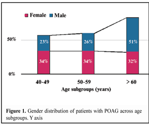
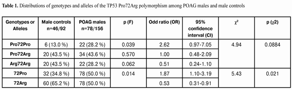
https://doi.org/10.31288/oftalmolzh201641519 Gender- and age-related features of the association between TP53 Pro72Arg polymorphism and primary open-angle glaucoma S.Iu. Mogilevskyy1, Dr Sc (Med), Prof. S.V. Ziablitsev2, Dr Sc (Med), Prof. L. I. Denisiuk1, Assistant 1Ophthalmology Department, Shupik National Medical Academy of Postgraduate Education, Ministry of Public Health of Ukraine 2Pathophysiology Department, Bohomolets National Medical University, Ministry of Public Health of Ukraine Odessa, Ukraine E-mail: sergey.mogilevskyy@gmail.com Background: The TP53 gene is directly related to the development of primary open-angle glaucoma (POAG). There have been inconsistent reports regarding increased risk of POAG and the C allele (72Pro) of the TP53 codon 72 polymorphism. Purpose: To identify associations, if any, between TP53 Pro72Arg polymorphism and presence of POAG in patients of different genders and age groups. Materials and Methods: The study group comprised 172 patients (78 men (45%) and 94 (55 %) women) diagnosed with POAG, and the control group comprised 98 individuals (46 men (47 %) and 52 women (53 %)) without POAG. The mean age of study participants was 57.3±1.1 years at the time of the examination for POAG. Results: In males with POAG, the frequencies of the 72Pro allele and of the Pro72Pro genotype of the ТР53 gene were found to be increased (1.4 times, р = 0.014 and 2.2 times, р = 0.039, respectively) compared to the relevant controls. The presence of the 72Pro allele in males (as well as in females) was associated with POAG. Male carriers of the 72Pro allele had a 1.87 (p (?2) = 0.021) times higher risk for the disease. Elevated frequencies of the 72Pro allele and the Pro72Pro genotype of TP53 codon 72 polymorphism were specific for POAG males older than 60 years, which had a 2.786 times higher risk for the disease (p (?2) = 0.019) compared to the relevant controls. Elevated frequency of the 72Pro allele, as well as decreased frequency of the 72Arg allele was more specific for POAG females younger than 60 years compared to the relevant controls. Female 72Pro allele carriers younger than 60 years had a 2.30 (p (?2) = 0.034) times higher risk for the disease compared to the relevant controls. Conclusions: The presence of the 72Pro allele in males was associated with POAG. POAG males older than 60 years had a 2.786 times higher risk for the disease compared to the relevant controls. The presence of the 72Pro allele in females was associated with POAG. Female 72Pro allele carriers younger than 60 years had a 2.30 times higher risk for the disease compared to the relevant controls. Key words: primary open-angle glaucoma, TP53 Pro72Arg polymorphism, gender, age Introduction Oxidative stress and apoptotic damage to the third retinal neuron play a special role in primary open-angle glaucoma (POAG), which is a multifactorial disease with a threshold effect [1]. Metabolic disorders, hypoxia and excessive IOP contribute significantly to the triggering of the apoptosis of retinal neurons [2]. Glaucoma affects individuals of both genders and different age groups. The incidence of POAG in males exceeds that in females by a factor of 3; however, low tension glaucoma prevails in women, with a women/men ratio of 2:1 [3, 4]. The prevalence of POAG has been reported [1] to be to as low as 0.1% in people 40-49 years of age, rising to as much as 14.3% in those over 80 years of age; the yearly incidence of glaucoma per 1,000 individuals aged 40 to 45 is approximately 1. The TP53 gene is one of the numerous hereditary and genetic factors resulting in the development of POAG, and is directly related to this development [5]. Since the gene codes the protein, p53, its expression is directly related to the implementation of apoptotic features of this protein. In recent years, as a potential candidate susceptibility gene for glaucoma, TP53 polymorphisms have attracted much attention in the medical community, and have been investigated in different populations. Although the number of known TP53 polymorphisms is more than 30, of the most significance for POAG is a single nucleotide polymorphism (SNP) in exon 4 of p53 at codon 72, a substitution of cytosine for guanine (Arg72Pro) [2, 5, 6]. The role of p53 in initiation of apoptosis is to transcriptionally activate proapoptotic proteins Вах, Noxa, p53AIP1 and Puma, which promote cytochrome c release from mitochondria and repression of antiapoptotic protein Bcl2 [6]. Another possible mechanism of the initiation of apoptosis induced by p53 involves an increase in intracellular free radical oxygen species due to activation of the genes involved in the regulation of cellular redox balance (PIG3, PIG8, FDXR) [6]. Therefore, in a neurodegenerative disease (particularly in POAG), apoptosis may result from cellular stress response and activation of the TP53 gene, and the genetic variability of this gene is of significant importance with regard to neuronal loss. Although some studies have suggested that the C allele (72Pro) [1, 3, 6, 8, 9], or, to the contrary, the proapoptotic allele G (72Arg) [2] of p53 codon 72 polymorphism is associated with POAG, other studies have reported on the lack of association between p53 gene polymorphisms and POAG [4, 9, 10, 11], which may depend on the age at which the disease manifests and between male and female patients. Therefore, of urgent importance is to elucidate the impact of TP53 polymorphism on individuals of different genders and age groups of Ukrainian population, given that in this population, such investigations have not yet been conducted. The study purpose was to identify associations, if any, between TP53 Pro72Arg polymorphism and presence of POAG in patients of different genders and age groups. Materials and Methods The study group comprised 172 patients diagnosed with POAG, and the control group comprised 98 individuals without POAG, the two groups being comparable in terms of female-to-male percentage (78 men (45%) and 94 (55 %) women versus 46 men (47%) and 52 women (53 %), respectively). The ages of study participants ranged from 40 to 74 years (mean, 57.3±1.1 years) at the time of the examination for POAG, the ages of men and women being 40 to 73 years (mean, 58.8±1.5 years) and 40 to 74 years (mean, 56.1±1.5 years), respectively. These indices were not statistically significantly different. Therefore, there was no statistically significant difference among the two groups in terms of age and gender. Study participants were subdivided into three age categories (low-age subgroup, less than 49 years; middle-age subgroup, 50 to 59 years; and high-age subgroup, more than 60 years) as per the WHO age classification scheme, and underwent visual acuity testing, Humphrey perimetry (as per World Glaucoma Association guidelines), pneumotonometry, autorefractometry, ophthalmoscopy and ocular coherence tomography (OCT), and changes in ocular hydrodynamics and visual function were investigated in controls at baseline, at one year and at the end of the study (at two years). Polymorphic variants of TP53 gene were determined by real-time polymerase chain reaction (PCR) using a TaqMan®SNP Genotyping Assay (Life Technologies, Grand Island, NY) and DTlite Real-Time PCR System (DNA Systems, Moscow, Russia). We estimated both the allele and the genotype frequencies of TP53 gene polymorphisms, and identified associations of the alleles and genotypes with disease status. After an informed consent was obtained, a 2.5-ml blood sample was drawn from the cubital vein and collected into the violet-capped Vacutainer System (Sarstedt, Germany) with a solution containing calcium ethylenediaminetetraacetic acid (EDTA; 11.7 mM) as an anticoagulant. Statistical analysis was performed using MedCalc v.15.11.0 (MedCalc Software bvba, 1993–2015). Contingency tables were used to test genotypes and alleles for association with the disease and to calculate odds ratios (OR) and 95% confidence intervals (CI). Fisher’s exact test was used to assess the statistical significance of frequency distributions. Pearson ?2 test was used to assess the likelihood of the impact of genotypes and alleles on disease status. Results and Discussion In the high-age subgroup, the percentage of male patients with POAG was higher than those in the middle-age ((р(F) = 0.008) and low-age subgroups ((р(F) = 0.001) (Fig. 1). There was no difference in the distribution of female patients with POAG among age subgroups. Therefore, POAG occurred more often in elderly males than in younger males, whereas the presence of the disease in females was not age-dependant. Given these findings, it was reasonable to perform separate comparisons of the allele and genotype distribution frequencies for TP53 Pro72Arg polymorphism among POAG males and male controls and among POAG females and female controls. The Pro72Pro genotype was found in 28.2% of males of the study group, which was 2.2 times more often than in males of the control group (13.0 %; p(F) = 0.039) (Table 1). There was no statistically significant difference in the distribution of other genotypes among males of the groups.
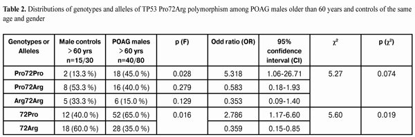
Therefore, we can assert that the frequency of Pro72Pro genotype of the TP53 gene in males with POAG was 2.2 times higher (р=0.039) than that in male controls. The distribution of alleles of the polymorphism under investigation was statistically different between males with POAG and male controls (Table 1). The frequency of the 72Pro allele in the study group (50%) was found to be 1.4 times as high as that in controls (34.8%). The 72Arg allele, on the contrary, was found significantly (1.3 times) less frequently in the main group than in controls (50.0% versus 65.2%; р(F) = 0.014). The distribution of alleles was found to be associated with POAG, with male carriers of the 72Pro allele having elevated (OR=1.87; CI=1.10-3.19; p (?2) = 0.021) risk for the disease. Male carriers of the 79Arg allele, correspondingly, had a 1.87 times decreased risk for the disease. Therefore, in the presence of POAG in males, the frequencies of the 72Pro allele and of the Pro72Pro genotype of the ТР53 gene were found to be increased (1.4 times, р=0.014 and 2.2 times, р=0.039, respectively). The presence of the 72Pro allele in males was associated with POAG, and male carriers of the allele had a 1.87 times higher risk for the disease (p (?2) = 0.021) compared with male non-carriers. Given significant age- and gender-related differences in the incidence of POAG, we decided to identify the associations, if any, between TP53 Pro72Arg polymorphism and presence of POAG among males younger than 60 and males older than 60, and among females younger than 60 and females older than 60. No statistically significant difference in the distribution of genotype and allele frequencies was observed between POAG males and male controls younger than 60 years. However, there was a difference in the distribution of genotype and allele frequencies between POAG males and male controls older than 60 years (Table 2).
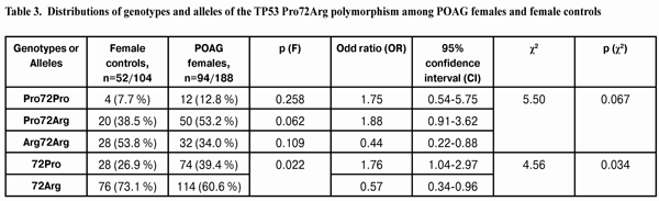
Thus, the frequency of the Pro72Pro genotype in POAG males older than 60 years (45%) was found to be 3.4 times as high as that in male controls older than 60 years (13.3 %; p(F) = 0.028). No significant difference in the distribution of other genotypes was found between POAG males and male controls older than 60 years. Therefore, the phenomenon demonstrated above (that of a higher frequency of the Pro72Pro genotype in POAG group males compared to that in control group males) was related mostly to males older than 60 years. That is, we can assert, that, in the presence of POAG in males aged only 60 years and more, the frequency of the Pro72Pro genotype in POAG males was 3.4 times (р=0.028) as high as that in male controls of the same age. In addition, a statistically significant difference in allele distribution was observed between POAG males and male controls older than 60 years (Table 2). The 72Pro allele was 1.6 times as common in POAG males older than 60 years than in controls of the same age (65.0% versus 40.0%). The 72Arg allele was found significantly (1.7 times) less frequently in POAG males older than 60 years than in controls of the same age (35.0% versus 60.0%; р(F) = 0.016). The allele distribution was found to be associated with POAG, and male carriers of the 72Pro allele older than 60 years had a 2.786 times higher risk for the disease (CI=1.17-6.60; p (?2) = 0.019) compared with male non-carriers of the same age. Male 79Arg-allele carriers older than 60 years, correspondingly, had a 2.786 times lower risk for the disease compared with male 79Arg-allele non-carriers of the same age. Table 3 presents the results of the analysis of the allele and genotype distribution frequencies for TP53 Pro72Arg polymorphism among POAG females and female controls.
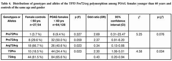
In POAG females, the Pro72Pro genotype was found 1.7 times more often than in female controls; however, this difference, contrary to that in males, was not of statistical significance (p (F) = 0.258). In addition, in POAG females, the Arg72Arg genotype was found 1.6 times less often than in female controls, and the difference was also not statistically significant (p (F) = 0.109). No significant difference in the frequency of heterozygote genotype Pro72Arg was found between POAG females and female controls (p (F) = 0.109). Correspondingly, no association was found between the TP53 Pro72Arg polymorphism and POAG in females (р (?2) = 0.067). There was a statistically significant difference in the distribution of the alleles (contrary to the distribution of the genotypes) of the polymorphism under investigation between POAG females and female controls (Table 3). The 72Pro allele was found significantly (1.5 times) more frequently in POAG females than in female controls (39.4% versus 26.9%; р(F) = 0.016). The 72Arg allele was found 1.2 times less frequently in study group females than in female controls (60.6% versus 73.1%; р(F) = 0.022). The distribution of alleles was found to be associated with POAG, i.e. female carriers of the 72Pro allele had elevated (OR=1.76; CI=1.04-2.97; p (?2) = 0.034) risk for the disease. Female carriers of the 79Arg allele had a 1.76 times decreased risk for POAG. Given significant age- and gender-related differences in the incidence of POAG, we decided to identify the associations, if any, between TP53 Pro72Arg polymorphism and presence of POAG among females younger than 60 and females older than 60. There was a difference in the distribution of genotype and allele frequencies between POAG females and female controls younger than 60 years (Table 4).
Thus, the frequency of the Pro72Pro genotype and the frequency of heterozygous genotype Pro72Arg in POAG females younger than 60 years were found to be 2.5 times and 1.7 times, respectively, as high as those in female controls of the same age, but these differences were statistically insignificant (p(F) = 0.327 and p (F) = 0.059, respectively). However, the frequency of the Arg72Arg genotype in POAG females younger than 60 years was found to be 1.6 times as low as that in female controls of the same age, and this difference was statistically significant (p(F) = 0.023). The distribution of genotypes among females younger than 60 years was not associated with POAG (р (?2) = 0.076). The distribution of alleles of the polymorphism under investigation was statistically different between POAG females younger than 60 years and female controls of the same age (Table 4). The 72Pro allele was found 1.9 times more frequently in study group females than in female controls (34.4% versus 18.5%, respectively). The 72Arg allele was found (1.2 times) significantly less frequently in study group females than in female controls (65.6% versus 81.5%, respectively; р (F) = 0.023). The distribution of alleles was found to be associated with POAG, i.e. female 72Pro allele carriers younger than 60 years had a 2.30 (CI=1.06-5.01; p (?2) = 0.034 ) times higher risk for the disease compared to the relevant controls. Female 79Arg allele carriers younger than 60 years, correspondingly, had a 2.30 times lower risk for the disease compared to the relevant controls. Analysis of the data obtained showed no significant difference in the distribution of genotype and allele frequencies between POAG females and female controls older than 60 years. Therefore, the phenomenon demonstrated above (that of a higher frequency of the 72Pro allele and a lower frequency of the 72Arg allele in POAG group females compared to those in control group females) was related mostly to females younger than 60 years. Conclusions First, in the presence of POAG in males, the frequencies of the 72Pro allele and of the Pro72Pro genotype of the ТР53 gene were found to be (1.4 times, р=0.014 and 2.2 times, р=0.039, respectively) increased. The presence of the 72Pro allele in males was associated with POAG, and male carriers of the 72Pro allele had a 1.87 times higher risk for the disease (p (?2) = 0.021). Analysis of the distributions of genotypes and alleles and of their associations with the development of POAG in males showed that, elevated frequencies of the 72Pro allele and the Pro72Pro genotype of TP53 codon 72 polymorphism was specific for POAG males older than 60 years, which had a 2.786 times higher risk for the disease (p (?2) = 0.019) compared to the relevant controls. Second, the presence of the 72Pro allele in females (similar to its presence in males) was associated with POAG. Analysis of the distributions of genotypes and alleles and of their associations with the development of POAG in females showed that, elevated frequency of the 72Pro allele, as well as decreased frequency of the 72Arg allele of TP53 codon 72 polymorphism was more specific for POAG females younger than 60 years compared to the relevant controls. Female 72Pro allele carriers younger than 60 years had a 2.30 (p (?2) = 0.034) times higher risk for the disease compared to the relevant controls.
References 1.Levkovitch-Verbin H, Vander S, Makarovsky D, et al. Increase in retinal ganglion cells susceptibility to elevated intraocular pressure and impairment of their endogenous neuroprotective mechanism by age. Mol Vis. 2013 Sep 26;19:2011-22 2.Logunov NA, Belousova AI, Vitkovskyy IuA. [The proapoptotic p53 (C72G) and p21 (C31A) polymorphisms as the risk factors for the development of primary open-angle glaucoma in Transbaikalian region]. Vestn Oftalmol. 2012 Sep-Oct;128(5):10-3 Russian 3.Lin HJ, Chen WC, Tsai FJ, Tsai SW. Distributions of p53 codon 72 polymorphism in primary open angle glaucoma. Br J Ophthalmol. 2002 Jul;86(7):767-70 4.Silva RE, Arruda JT, Rodrigues FW, Moura KK. Primary open angle glaucoma was not found to be associated with p53 codon 72 polymorphism in a Brazilian cohort. Genet Mol Res. 2009;8:268–272 5.Liu T, Lin X, Jian Y, Yuewuyang L, et al. Screening of candidate genes for primary open angle glaucoma. Mol Vis. 2012;18:2119-26 6.Chumakov PM. [Protein p53 and its universal functions in the multicellular body]. Advances in Biological Chemistry. 2007;47:3–52 Russian 7.Acharya M, Mitra S, Mukhopadhyay A, et al. Distribution of p53 codon 72 polymorphism in Indian primary open angle glaucoma patients. Mol Vis. 2002 Sep 30;8:367-71 8.Daugherty CL, Curtis H, Realini T, et al. Primary open angle glaucoma in a Caucasian population is associated with the p53 codon 72 polymorphism. Mol Vis. 2009 Sep;15:1939–44 9.Neamatzadeh H, Soleimanizad R, Zare-Shehneh M, et al. Association between p53 codon 72 (Arg72Pro) polymorphism and primary open-angle glaucoma in Iranian patients. Iran Biomed J. 2015;19(1):51-6 10.Fan BJ, Liu K, Wang DY, et al. Association of polymorphisms of tumor necrosis factor and tumor protein p53 with primary open-angle glaucoma. Invest Ophthalmol Vis Sci. 2010 Aug;51(8):4110-6 11.Mabuchi F, Sakurada Yo, Kashiwagi K, et al. Lack of association between p53 gene polymorphisms and primary open angle glaucoma in the Japanese population. Mol Vis. 2009;15:1045–1049 12.Saglar E, Yucel D, Bozkurt B, et al. Association of polymorphisms in APOE, p53, and p21 with primary open-angle glaucoma in Turkish patients. Mol Vis. 2009;15:1270–1276
|