J.ophthalmol.(Ukraine).2016;2:41-43.
|
https://doi.org/10.31288/oftalmolzh201624143 Membrane trophic effect of hyperglycemia on the anterior chamber angle tissues in ocular hypertension V.R. Yurevich, Cand. Sc. (Med), Ass. Prof. Danylo Halytsky Lviv National Medical University Lviv (Ukraine) E-mail: yurevych@yahoo.com Introduction. Although there is a significant progress in the treatment of diabetic ocular complications, the issue of prevention and therapy for this pathology is still unstudied. The purpose of the present study was to study the stability of lysosomal membranes in the anterior chamber angle tissues in ocular hypertension under hyperglycemia conditions. Materials and Methods. A total of 32 male chinchilla rabbits, weight 2.5-3.2 kg, were involved into the experiment.The experimental animals were divided into 4 groups: group I, 8 rabbits served as control; group II, 8 rabbits with DB under ocular hypertension conditions (DM+OH group); group III, 8 rabbits with DM (DM-only group); group IV, 8 rabbits with ocular hypertension (OH-only group). Each group was subdivided into 2 subgroups depending on the time point of the follow up: subgroup i, 3 weeks; subgroup ii, 6 weeks. Activity of free and bound forms of acid phosphatase (ACP) in the anterior chamber angle tissues was determined. Results. Data obtained revealed that ocular hypertension development in streptozotocin-induced diabetic rats caused significant decrease in stability of lysosomal membrane in intracellular structure of tissues studied. Conclusions. It was established that under the conditions of ocular hypertension, streptozotocin-induced diabetic animals had a marked failure in the stability of anterior chamber angle tissue lysosomes. In such conditions, the activity of free marker enzyme increased by more than 40% and the level of bound marker enzyme decreased by 47.7%. Whereas, these indices were equal to 26.1% and 25.3%, respectively, when ocular hypertension was simulated in normoglycemic rabbits. Thus, we can conclude that hyperglycemia has a negative effect on the intracellular ultra structures of the anterior chamber angle tissues in the development of ocular hypertension. Key words: ocular hypertension, diabetes, anterior chamber angle tissue, acid phosphatase
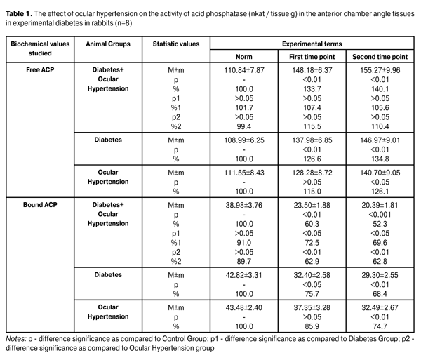
Introduction The issue related to the search of new methods for preservation of visual functions in primary glaucoma is of great importance today and it becomes even more relevant when treating glaucoma in diabetic patients [1, 7]. Glaucoma and diabetes mellitus (DM) have been suggested to have the similarity in pathogenic mechanisms. In particular, when DM develops, the role of oxidative stress has been defined in the mechanisms of diabetic damage of the eye [3, 7]. There have also been reports on the role of oxidative processes in the pathogenesis of glaucomatous optic neuropathy [4]. Meanwhile, it is still unclear how activation of lipid peroxidation (LPO) is correlated to the state of the antioxidant system in the blood, eye drainage system tissues and aqueous humor in primary open-angle glaucoma (POAG) [2, 9]. And to our best knowledge, the character of free radical damage of membrane structures of anterior chamber angle tissues in POAG under DM conditions has been unstudied. We believe that complex investigation of LPO and antioxidant protection processes as well as the structural and functional state of lysosomes in POAG and DM is of great interest not only for improving the knowledge in the disease-related pathogenesis but for development of new pharmacology methods for these diseases. The purpose of the present study was to study the stability of lysosomal membranes in the anterior chamber angle tissues in ocular hypertension under hyperglycemia conditions. Material and Methods A total of 32 male chinchilla rabbits, weight 2.5-3.2 kg, were involved into the experiment. All work with animals followed Guiding Principles for Biomedical Research Involving Animals issued by Council for the International Organizations of Medical Sciences (2012). The experimental animals were divided into 4 groups: group I, 8 rabbits served as control; group II, 8 rabbits with DM under ocular hypertension conditions (DM+OC group); group III, 8 rabbits with DM (DM-only group); group IV, 8 rabbits with ocular hypertension (OH-only group). Each group was subdivided into 2 subgroups depending on the time point of the follow up: subgroup i, 3 weeks; subgroup ii, 6 weeks. The models for simulating DM and ocular hypertension (OH) used as well as the animal euthanizing have been described in our previous papers [5, 6]. Tissues studied were immediately removed and placed in as-prepared medium, containing 20 мМ HEPES-KOH (pH=7.5), 1.5 M MgCl2, 0.5 mM EGTA and 250 mM sucrose; afterwards, they were homogenized with a Teflon pestle in a glass homogenizer. After differential centrifugation of homogenates in pellet and supernatant fluid, activity of free and bound forms of acid phosphatase (ACP) was determined [4]. A method of determining the ACP activity is based on the spectrophotometric (CP) assay of concentration of para-nitrofenyl which had been formed in a result of enzymatic hydrolysis of para-Nitrophenylphosphate. The coefficient of variation of the technique was 3.6% [8]. SPSS student software was used to determine statistical significance of differences [8]. Results and Discussion Data on the effect of hyperglycemia on the activity of free and bound ACP in the anterior chamber angle tissues in rabbits with OH are given in Table 1. Correlation of free and bound ACP activities was considered to assess the degree of lysosoma labilizing. Activity of free ACP in the anterior chamber angle tissues in OH rabbits increased up to 115.0% and 126.1% at the first and second time points, respectively, as compared to norm. Activity of free ACP in the anterior chamber angle tissues in DM and OH rabbits was increased up to 133.7% (р<0.01) and 140.1%, at the first and second time points, respectively, as compared to the control (р<0.01). Obviously, activity of free ACP increased more significantly in rabbits with DM and OH than in non-diabetic rabbits induced OH. Activity of bound ACP in the anterior chamber angle tissues in OH rabbits decreased to 85.9% and 74.7% at the first and second time points, respectively, as compared to norm (р<0.01).
Activity of bound ACP in the anterior chamber angle tissues in DM and OH rabbits was decreased to 60.3% (р<0.01) and 52.3%, at the first and second time points, respectively, as compared to the control (р<0.001). At time points studied, a significant fall in activity of bound ACT was noted in DM and OH rabbits as compared to DM-only animals. Thus, the decrease was equal to 27.5% (р<0.05) and 30.4% (р<0.05) at the first and second time points, respectively. It should be pointed that activity of bound ACP decreased in much more degree in DM and OH rabbits than in non-diabetic rabbits induced OH. Thus, a relative decrease was equal to 37.1% and 37.2% (р<0.01) at the first and second time points, respectively. Totally, the general analysis of the presented findings of studying the activity of free and bound form of marker enzyme ACP indicates to the fact that OH development in streptozotocin-induced diabetic rabbits results in significant decrease in stability of lysosomal membranes of intracellular structures in the tissues studied. The general analysis of the experimental data obtained from different group animals makes it possible to conclude that OH simulation under the conditions of hyperglycemia leads to the more pronounced disorders in lysosoma stability as compared to a simulation model when OH was induced in rabbits with normal glycemia. Activation of oxygen free radical formation process, an increase in LPO and a decrease of antioxidant system and enzyme detoxication potential can play an important role in the mechanisms of revealed disorders of membrane stability of the intracellular structures in the anterior chamber angle tissues; this fact was shown in our previous papers [5, 6]. Lyssomal stability is the important part in the destruction of the trabecular meshwork in the development of glaucomatous process since the state of these intracellular organelles determines the hydrolysis rates of trabecular meshwork macromolecules (protein, mucopolysaccharide etc.). Thus, the facts of lysosomal membrane stability disorders revealed in our work are of great importance [9]. Besides, it should be also considered that destabilization of lysosomal membranes is an important element in the apoptosis of cellular structures since these are lysosomal enzymes that provide catabolism of protein, lipid and other biopolymers. Conclusions It was established that under the conditions of ocular hypertension, streptozotocin-induced diabetic animals had a marked failure in the stability of anterior chamber angle tissue lysosomes. In such conditions, the activity of free marker enzyme increased by more than 40% and the level of bound marker enzyme decreased by 47.7%. Whereas, these indices were equal to 26.1% and 25.3%, respectively, when ocular hypertension was simulated in normoglycemic rabbits.
Thus, we can conclude that hyperglycemia has a negative effect on the intracellular ultra structers of the anterior chamber angle tissues in the development of ocular hypertension. References
|