J.ophthalmol.(Ukraine).2016;1:8-11.
|
https://doi.org/10.31288/oftalmolzh20161811 Visual function status in myopic patients before and after pharmacological correction of antioxidant defense system failure E.I. Surova I.M. Boichuk, Dr. Sc. (Med) Filatov Institute of Eye Diseases and Tissue Therapy Odessa, Ukraine E-mail: iryna54@mail.ru Background: Recent studies of the clinical features and pathogenesis of the acquired, progressive and complicated myopia have evidenced that patients with this disease have reduced antioxidant defense. It is hypothesized that pharmacological correction of antioxidant defense system failure will improve visual functions in myopes. Purpose: To assess the status of visual functions (including distance visual acuity, absolute reserve of accommodation and photopic light sensitivity) in patients with myopia of different degrees before and following treatment with an antioxidant medication (Fakovit). Materials and Methods: The Fakovit treatment group included 70 myopes (140 eyes) (mild myopes, n = 28; medium myopes, n = 22; high myopes, n = 20), whereas the control group included 30 aged matched myopes who did not receive Fakovit treatment. All patients underwent ophthalmic examinations including refractometry, biomicroscopy, echobiometry, ophthalmoscopy, and absolute reserve of accommodation. The uncorrected and corrected distance visual acuities (VA) were assessed using the Shevalev’s Chart, and absolute reserve of accommodation (RA) was assessed using a conventional technique. Photopic light sensitivity was assessed with the use of semi-automatic registering adaptometer. Results: An improvement in VA was achieved in 86.1% of mild and medium myopes of the treatment group. An improvement in and stabilization of RA was found in 44.4% (mainly, in mild myopes) and 38.8%, respectively, of the patients of the treatment group, and a 1.43±1.29-D reduction in RA (p < 0.05) was observed in the control group. Photopic light sensitivity of the visual system had a tendency to increase in all myopes after treatment with Fakovit (in particular, it increased statistically significantly by 0.16±0.04 log units in mild myopes), and did not change in the controls. Conclusion: Correction of antioxidant defense system failure with Fakovit improved and stabilized visual functions (visual acuity, reserve of accommodation, and photopic light sensitivity) in myopic patients. Key words: pharmacological correction, antioxidant defense system, reduced antioxidant defense, myopia.
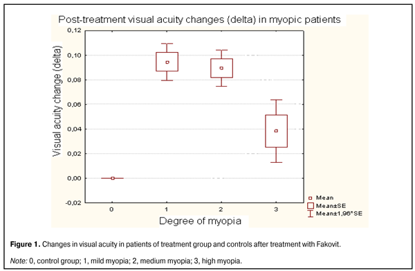
Introduction Myopia is still one of the most important problems of ophthalmology. The prevalence of the disease has been increasing globally and it has been estimated that 30% to 60% of the worldwide population are myopes [1]. Recent studies of the clinical features and pathogenesis of the acquired, progressive and complicated myopia have evidenced activation of lipid peroxidation (LPO) in metabolism of ocular tissues and reduced antioxidant defense in myopic patients [1-8]. It is known that free radicals can affect cell membranes, and antioxidant mechanisms of the body can protect the latter from oxidative stress. In normally functioning systems of the body, there is an adequate relationship between lipid peroxidation activity and antioxidant defense level. Given the fact that, in myopia, one may observe not only a reduction in the main functions of the visual system, but also impaired biochemical and immunological indices, scleral alterations, and increased lipid oxidation, current medical treatment of myopia involves different medications [6-10]. In recent years, there have appeared studies of one of the most powerful endogenous (intracellular) antioxidants, glutathione, and the possibility for increasing the levels of antioxidant thiol glutathione through the introduction of antioxidant medications (Coenzyme Compositum, in particular) into the therapy for dry age-related macular dystrophy [11]. That is why our attention has been drawn to Fakovit (an analog of Fakolen), an antiradical and detoxification agent which has been successfully used in the treatment of cataracts and lens metabolism disorders. Fakovit (pharmaceutical corporation Zdorovie, Kharkiv, Ukraine) is available in tablets composed of glutamic acid, glycine, pyridoxine hydrochloride, L-cysteine and ascorbic acid. Being an endogenous antioxidant, glutathione, a major component of the thiol-disulphide system, is a tripeptide derived from glutamic acid, glycine and cysteine intracellularly through the action of adenosine triphosphoric acid. The main functions of this tripeptide are protection of thiol proteins against oxidative damage, quenching of free radicals, and elimination of foreign organic compounds. Amino acid transport through membranes is performed through the action of phosphorylated form of pyridoxine hydrochloride (pyridoxal phosphate). Ascorbic acid regulates intracellular redox processes and is involved in water exchange and restoration of disulfide bonds. Our previous study has demonstrated that the treatment of myopes with Fakovit was followed by an increase in antioxidant blood levels [12]. Therefore, the purpose of this study was to assess the status of visual functions (including distance visual acuity, absolute reserve of accommodation, and photopic light sensitivity) in patients with myopia of different degrees before and following treatment with Fakovit. Materials and Methods The Fakovit treatment group included 70 myopes (140 eyes) (mild myopes, n = 28; medium myopes, n = 22; high myopes, n = 20) aged 12-24 years, whereas the control group included 30 aged-matched myopes (mild myopes, n = 20 eyes; medium myopes, n = 20 eyes; high myopes, n = 20 eyes) who did not receive Fakovit treatment. All patients underwent ophthalmic examinations including refractometry, biomicroscopy, echobiometry, ophthalmoscopy, and absolute reserve of accommodation. The uncorrected and corrected distance visual acuities at 5 m were assessed using the Shevalev’s Chart, and absolute reserve of accommodation was assessed using a conventional technique [1]. Light sensitivity (LS) of the visual system was assessed with the use of semi-automatic registering adaptometer (SRA) operating in specified mode (3 minutes of light adaptation followed by 60 minutes of dark adaptation) and through an absolute visual threshold determined during dark adaptation. Each eye was examined monocularly. Visual light sensitivity threshold in dark adaptation mode was determined every two minutes for 10 minutes (cone cell stage or photopic light sensitivity threshold). Visual functions were assessed before and 1-1.5 months after treatment with Fakovit. Fakovit treatment regimen was as follows: a tablet of each type (those dissolved in the stomach and those dissolved in the gut) twice a day with meals within a 30-day treatment course, four courses a year. STATISTICA 8 for Windows software package was used for statistical analysis with an analysis of variance. Results Figure 1 shows the visual acuity data in patients with different degrees of myopia before and after treatment with Fakovit. After treatment, visual acuity in mild and medium myopes of the treatment group improved, whereas those in high myopes of the treatment group and myopic controls did not improve significantly. In mild and medium myopes of the treatment group, the mean improvement in VA was 0.09±0.05 units and 0.08±0.03 units, respectively, which was statistically significantly more than in the control group. Therefore, the improvement in VA was achieved in 86.1% of mild and medium myopes.
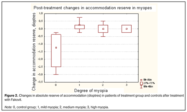
After treatment with Fakovit, a 0.25 D to 0.75 D improvement, no change, and a 0.25 to 0.5 D reduction in reserve of accommodation (RA) was observed in 44.4%, 38.8%, and 16.6%, respectively, of patients of the treatment group. At the same time, a 1.43±1.29 D reduction in RA (p<0.05) was observed in all controls (Fig. 2).
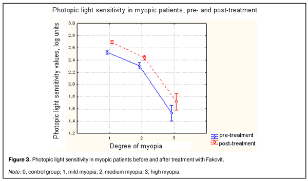
Figure 3 shows the results of the assessment of photopic light sensitivity of the visual system before and after treatment with Fakovit.
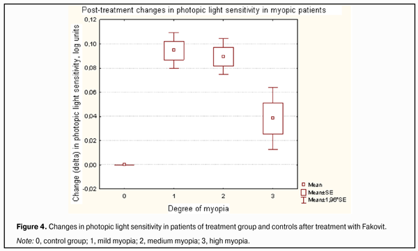
It is seen that after treatment with Fakovit, the photopic light sensitivity improved most of all in mild myopes (0.16±0.04 log units) followed by medium myopes (0.13 ± 0.06 log units) and high myopes (0.13 ± 0.08 log units) of the treatment group, but not significantly. However, the photopic light sensitivity in all patients of the treatment group increased significantly compared to controls (р < 0.05; Fig. 4).
Conclusion Correction of antioxidant defense system failure with Fakovit, an antioxidant medication, improved and stabilized visual functions (visual acuity, reserve of accommodation, and photopic light sensitivity) in myopic patients. An improvement in VA was achieved in 86.1% of mild and medium myopes of the treatment group. Additionally, an improvement in and stabilization of RA was found in 44.4% (mainly, in mild myopes) and 38.8%, respectively, of the patients of the treatment group, and a 1.43±1.29-D reduction in RA (p < 0.05) was observed in the control group. Finally, photopic light sensitivity of the visual system had a tendency to increase in all myopes after treatment (in particular, it increased statistically significantly by 0.16±0.04 log units in mild myopes), and did not change in the controls. References 1. Avetisov ES. [Myopia]. Moscow: Meditsina; 1986. 240 p. Russian 2. Bushuieva NN. [Current aspects of etiology, pathogenesis and surgical treatment of progressive myopia]. Oftalmol Zh. 2006;3(1):65-70. Russian 3. Vladimirov IuA, Archakov AI. [Lipid peroxidation in biological membranes]. Moscow: Nauka; 1970. 252 p. Russian 4. Vinetskaia MI, Kushnarevich NIu, Tarutta EP, Demchuk ML. [Investigation of indices of tear oxidation by free radicals in progressive myopia]. In: [Current Issues in Ophthalmology. Proceedings of the Conference on the 170th Anniversary of Moscow City Ophthalmological Clinical Hospital]; 1996; Moscow; p.51-2. Russian 5. Kozlov IuP. [Structural and functional aspects of lipid peroxidation in biological membranes]. In: [Lipids: structures, biosynthesis, transformations and functions. Collection of scientific papers.] p.80-93. Russian 6. Stotska LM, Serdiuchenko VI. [Efficiency of spirulina bio-regulator in the treatment of uncomplicated acquired myopia in children and adolescents]. Oftalmol Zh. 2005;3:36-40. Ukrainian 7. Tarutta EP. [Potential for prevention of progressive and complicated myopia in light of current knowledge on pathogenesis of this disease]. Vestn Oftalmol. 2006;1:43-7. Russian 8. Metelitsyna IP, Stotskaia LM, Serdiuchenko VI. [Efficacy of spirulina in treatment of myopia in children based on biochemical and clinical indices]. Oftalmol Zh. 2004;6:54-8. Russian 9. Rykov SO. [Prevention of ocular disorders in children. Guidance manual]. Kyiv: SPT Bavok; 2003. 64 p. Ukrainian 10. Lee J, Lee HK, Kim CY et al. Purified high-dose anthocyanoside oligomer administration improves nocturnal vision and clinical symptoms in myopia subjects. Br J Nutr. 2005;93(6):895-9. 11. Iurevych OIu. [Clinical and experimental basis for correction of impaired thiol metabolism in patients with age-related macular dystrophy]. [Abstract for Cand. Sc. (Med) Thesis]. Odessa: Filatov Institute of Eye Diseases and Tissue Therapy; 2005. 16 p. Ukrainian 12. Surova EI, Boichuk IM, Kolomiichuk SG. [Status of blood antioxidant enzyme system in patients with different degrees of myopia before and after pharmacological correction of system failure]. Oftalmol Zh. 2014;6:34-9. Russian
|