J.ophthalmol.(Ukraine).2016;1:55-57.
|
https://doi.org/10.31288/oftalmolzh201615557 Restorative potential of eye lens glutathione system of animals with experimental ocular hypertension at light illumination N.F. Leus, Dr. Sc. (Med), Prof M Aldakhdukh, PhD student Yu. A. Zhuravok, Cand. Sc. (Med) Filatov Institute of Eye Diseases and Tissue Therapy Odessa, Ukraine E-mail: nusha_87@inbox.ru Introduction. The question of age-related cataract treatment effectiveness is one of the most important in ophthalmology. This question becomes particularly topical if this disease is progressed together with glaucomatous process. Purpose. To study the condition of glutathione system of animals eye lens with experimental ocular hypertension while modeling age-related cataract with the help of light illumination. Methods. Experimental studies were carried out on 39 rabbits, which were divided into three groups: in first group (9 animals) the light cataract was modeled, in the second group (8 animals) ocular hypertension was modeled, and in third one (10 animals) the ocular hypertension was induced after light illumination. Control group comprised 12 animals. In eye lenses and chamber liquid of experimental animals, the levels of reduced and oxidized glutathione were detected. Results. After general estimation of this study results it can be stated, that under the influence of cataractogenic factor, light energy, more sharp drop of restorative potential of glutathione in eye lenses was indicated in conditions of ocular hypertension as compared with studies, in which the light illumination was influenced in conditions of normal intraocular tension. Conclusion. It was indicated that modeling of light cataract in conditions of ocular hypertension causes more sharp injure of eye lens glutathione system as compared with cataractogenic influence of just light energy. The data on the high level of glutathione restorative potential reduction discovered an important link of cataractogenic influence of ocular hypertension and gives ground to consider it to be a risk factor of cataract progression Key words: age-related cataract, ocular hypertension, eye lens, glutathione
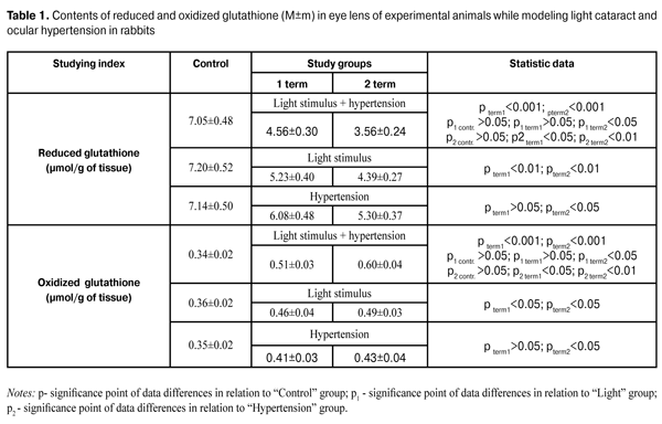
Introduction The issues on improvement of treatment effecacy of age-related cataract have become more topical. Especially important this problem is when this disease is progressed together with glaucomatous process [1]. The latter is considered as risk factor for cataract progression [3,8]. Due to studies, which have been carried out for the last two decades, the role of oxidation reduction system of glutathione in the process of cataractogenesis has been disclosed [7]. The abnormalities level of proteins thiolic groups and glutathione in the retina and ophthalmic nerve at development of glaucomatous process have been also detected [6]. At the same time, the condition of glutathione system in eye lenses and chamber liquid with progressing of age-related cataract of glaucoma patients remains unstudied. The purpose of the present paper was to study the condition of glutathione system of animals eye lenses with experimental ocular hypertension while modeling age-related cataract with the help of light illumination. Material and Methods The study was carried out on 39 Chinchilla male rabbits (weight, 2.5 – 3.2 kg), including 12 rabbits of control group. The study was carried out according to International principles of biomedical studies on animals, proposed by Council for International Organizations of Medical Sciences (CIOMS) (2012). General study scheme has been described previously [3]. The intraocular pressure averagely was (15.2±0.7) mm of mercury and (26.5±1.4) mm of mercury in control and study groups, respectively. The first group: 9 rabbits were modeled with light cataract; the second group: 8 rabbits were induced ocular hypertension; the third group: 10 rabbits were diseased with ocular hypertension before light illumination. The method of modeling of light cataract was described in the previous works [2,4]. To induce ocular hypertension, 0.1 ml of 0.3% solution of carbopol was injected in the anterior chamber of animal eye [9]. Collection of samples for study was carried out after 5 and 10 weeks (1 and 2 terms). In eye lens and chamber liquid of experimental animals, the contents of reduced and oxidized glutathione was spectrofluorimetrically detected according to abovementioned methods [2]. Data processing was carried out using SPSS software 11.0 [5]. Results and Discussion The level of reduced glutathione in eye lenses of studied animals with cataract and ocular hypertension deacreased at all terms of studyingas compared to controls, i.g. by 35.5%, and 45.5%, at terms 1 and 2, respectively. It is necessary to note that the level of reduced glutathione of animals with cataract and ocular hypertension were considerably lower than that of animals with cataract without ocular hypertension. Thus, it was 12.8% and 18.9%, at 5 and 10 weeks, respectively (Table 1).
At the same time, the level of oxidized glutathione of studied animals with cataract and ocular hypertension increased during the whole period of study by 50.0%and 76.5%, at term 1 and in 2, respectively, comparing to the control group. Besides, the level of oxidized glutathione of studied animals with progressed ocular hypertension was considerably higher than in groups of animals without ocular hypertension. So, in the first and second terms, this increasing was 10.9% and 22.4%, respectively (Table 1). The level of reduced glutathione in chamber liquid of animals with cataract and ocular hypertension decreased by 29.6% and 43.8% at the first term and second terms, respectively, as compared to norm (1.62±0.09) µmol/l, and it is necessary to note, that the level of reduced glutathione in chamber liquid of animals with cataract and ocular hypertension decreased more than that in chamber liquid of animals with cataract without ocular hypertension, i.g. by 9.8% and 20.2% at the first and second terms, respectively (р<0.05). While estimating generally the results obtained in this study, it can be stated than under the influence of light energy cataractogenic factor, more sharp decreasing of reduced potential of glutathione in eye lens ocures under the conditions of ocular hypertension as compared with experiments where light illumination was carried out under the conditions of normal intraocular tension. Considering the fact that the reduced potential of glutathione is a basic shielding component of eye lens from exposure of cataractogenic factors, this fact of more considerable damage of reduction system of glutathione in combination of light illumination and ocular hypertension can be regarded as cataractogenic influence of ocular hypertension to the process of light cataract progressing. Thus, obtained experimental data about damage of glutathione status uncover an important part of the mechanism of accelerated progressing of damage of eye lens optical characteristics, which was stated by using the combination of light energy and ocular hypertension in the previous study [3]. Conclusions 1.It was indicated that modeling of light cataract in conditions of ocular hypertension caused more sharp injure of eye lens glutathione system as compared with cataractogenic influence of light energy in conditions of normal intraocular tension. 2.The data on the high level of glutathione reduction potential discover an important link of cataractogenic influence of ocular hypertension and give ground to consider it to be a risk factor for cataract progression. References 1.Kurisheva NI, Fedorov АА, Yerichev VP. [Pathomorphological peculiarities of cataract eye lens of glaucoma patients]. Vestnik Oftalmologii. 2000;116(2):13-16. Russian. 2.Leus NF, Budaya Nizar, Girzheva АV. [Mechanizm of anticataractogenic action of carotinoids and flavonoids]. Oftalmologiia. Vostochnaya Evropa. 2013;3:86-94. Russian. 3.Leus NF, Aldakhdukh M. [Peculiarities of progressing of experimental form of age-related cataract in conditions of ocular hypertension]. Oftalmol Zh. 2015;3:91-95. Russian. 4.Leus NF, Metelitsina IP, Drozhzhina GІ and others. [Modeling method of radiation cataract]: Pat. 20178 Ukraine, PMK G 09 В 23/28, № 4712831/SU; Application 13.07.89; Published 25.12.97; Bul. “Prom. Vlasn.”;6:2–576. Russian. 5.Rebrova ОY. [Statistical analysis of medical data. Usage of the package of application programs STATISTICA]. M.: Media Sphera. 2002:312. Russian. 6.Serdyuk VN. [Studying of restorative potential of glutathione in retina and ophthalmic nerve while modeling of wide-angle glaucoma of rabbits]. Tavricheskii mediko-biol. vestnik. 2011;14:142-145. Russian. 7.Chamberlain CG, Mansfield KJ, Cerra A. Glutathione and catalase suppres TGF?-induced cataract-related changes in cultured rat lenses and lens epithelial explants. Mol. Vis. 2009;15:895-905. 8.Chandrasekaran S, Cumming RG. Association between elevated intraocular pressure and glaucoma, use of glaucoma medications, and 5-year incident cataract: the Blue Mountains Eye Study. Ophthalmol. 2006;113:417-424. 9.Wang YY. Experimental study of carbomer glaucoma model in rabbits by injecting different location in anterior chamber. Ophthalmol. 2009;45:1-95.
|