J.ophthalmol.(Ukraine).2016;1:51-54.
|
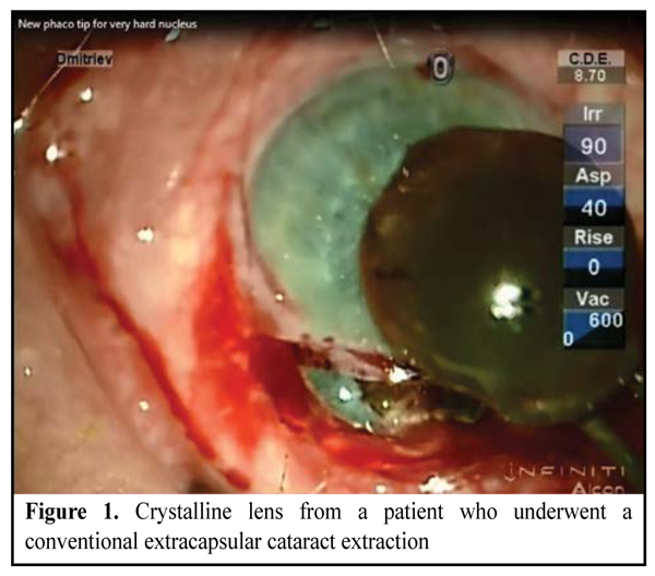
https://doi.org/10.31288/oftalmolzh201615154 Features of new phaco needle model for penetration of lens nucleus: an experimental study Ya.A. Gritsenko, MD S.K. Dmitriev, Dr. Sc. (Med), Prof N.V. Pasyechnikova, Dr. Sc. (Med), Prof Filatov Institute of Eye Diseases and Tissue Therapy Odessa, Ukraine E-mail: awsed2005@ukr.net Background. Although ultrasound phacoemulsification (phaco) is a common method for removing a cataract for different lens densities, the phaco of a high-density nucleus remains an important issue. This is primarily associated with the technical difficulties of removing the high-density nucleus when it is impossible to break it up completely. Purpose. To determine and compare the forces required for improved and conventional phaco needles to penetrate the isolated dense crystalline lens in vitro. Materials and Methods. The experimental study involved 15 crystalline lenses from patients who had undergone a conventional extracapsular cataract extraction. Two phaco needles, a conventional phaco needle (INTREPID® Micro-Coaxial System) and an improved one (Patent of Ukraine for the useful model No. 64851), were used in experiments. Results. In the in vitro experimental study, it was established that the mean force measurement value for the improved phaco needle was 1.4 times lower than that for the conventional needle. Conclusion. The use of the improved needle for the phaco of a high-density nucleus is reasonable and makes it possible to penetrate deeply into the lens and divide the lens into fragments in a prompter and more efficacious manner than with the use of the conventional phaco needle. Key words:improved phaco needle, phacoemulsification Introduction Although ultrasound phacoemulsification (phaco) is a common method for removing a cataract for different lens densities, the phaco of a high-density nucleus remains an important issue. Ioshin et al (2000), Kopaieva (2002) and Malyugin (2002) reported that high lens density and weak Zinn ligaments can complicate the course of surgery, with increased cumulative dissipated energy and aspiration time, and can result in intra- and postoperative complications [1]. This is primarily associated with the technical difficulties of removing the high-density nucleus when it is impossible to break it up completely. Hardness is the ability of the material to resist the penetration of an indenter under the action of a certain force. Experimental findings have proved that there is an apparent relationship between nuclear hardness and color and grade of nuclear sclerosis [2-3]. A relationship between optical and mechanical characteristics of the lens has been investigated in a number of studies. Thus, specially designed devices, lens guillotines and punches, were used to study the hardness of cataract nuclei from patients who had had extracapsular cataract surgery [4-6]. Korosteleva and Neresov (1990) developed an objective method to assess the lens hardness taking into account nuclear color and patient’s age [3]. The method involved the use of a dynamometer with a probe penetrating the lens at a specified penetration speed. Chuprov (2001) used a probe penetration technique to determine nuclear hardness, with the difference in the depth of indenter penetration into the sample under the action of preliminary and total loads measured experimentally. Avetisov (2011) developed a test bench to assess viscoplastic properties and fragility of biological tissues quantitatively under experimental conditions. He found a direct correlation between lens echogenicity and viscoplastic properties of the isolated lens [1]. Determination of forces required for phaco needles of different patterns to penetrate the lens is an important issue, given the reports on the possibility to conduct experimental studies for the assessment of mechanical properties of the lens. The purpose of the study was to determine and compare the forces required for improved and conventional phaco needles to penetrate the isolated dense crystalline lens in vitro. Materials and Methods The experimental study invo lved 15 crystalline lenses from patients who had undergone a conventional extracapsular cataract extraction. The experiments were conducted within 3 hours of harvesting the material (Fig. 1). Patients’ age varied from 56 to 82 years. Patients underwent assessment of the level of visual acuity, biomicroscopy of the anterior eye under conditions of pharmacological mydriasis, determination of lens density and ultrasound B scanning of the lens. Lens density measurements were verified by preoperative ultrasound B scanning with determination of relative acoustic impedance of different lens layers.
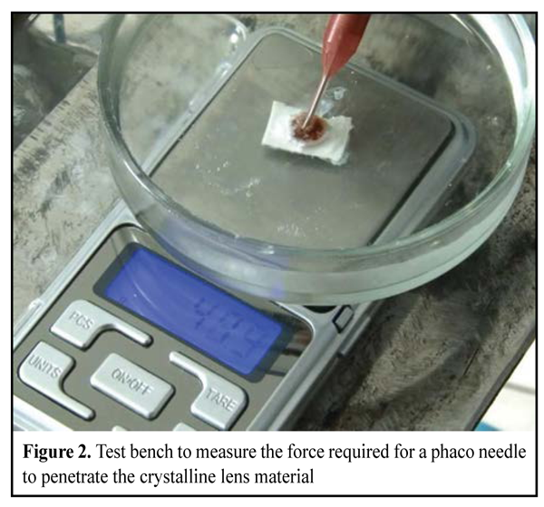
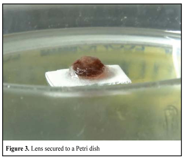
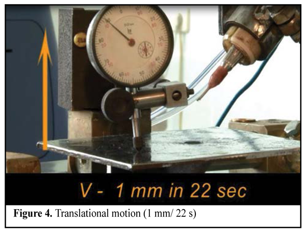
Two phaco needles, a conventional phaco needle (INTREPID® Micro-Coaxial System) and an improved one (Patent of Ukraine for the useful model No. 64851), were used in the experiments. The improved phaco needle has the following feature when compared with the conventional one: the end surface of the flared portion has a plurality of isosceles teeth, allowing the needle to penetrate deep layers of a high-density nucleus [9-10]. The samples were stored in balanced salt solution at 5—7°С. We have developed a test bench to measure the force required for a phaco needle to penetrate the crystalline lens material (Fig. 2). The test object was secured to the bottom of a Petri dish (Fig. 3). The dish with the lens was set on an electronic analytical balance with a readability of 0.01g. The INFINITI phaco handpiece was secured in a special clamp. The dish with the lens was fixed to a movable arm of the original device in a way ensuring its linear displacement towards the secured phaco handpiece. The speed of the movable arm was constant (1 mm/ 22 s) (Fig. 4).
Measurement of the force required for a phaco needle to penetrate the nucleus was read from the balance display at the time when the lens was contacted by a phaco needle operating at 100 percent longitudinal ultrasound and 100 percent torsional ultrasound. The duration of the experiment (45 s) was long enough to allow the needle to achieve the densest structures of the nucleus. The method consisted in measuring the force required for a phaco needle to penetrate into the nucleus. Force measurements were taken at the time of direct needle contact with the lens surface and on achieving maximum force value. Results and Discussion Nuclei of the study were divided into two groups based on nuclear color grades and absence of red fundus reflex, with Group 1 and Group 2 comprising 6 grade 4 nuclei and 9 grade 5 nuclei, respectively (Buratto classification scheme). Since the ultrasound examination revealed that the relative acoustic impedance of the lenses was 0.35 to 0.42 units, these lenses can be classified as mechanically hard. The mean lens thickness in Group 1 and Group 2 was 4.14 ± 0.16 mm, and 4.37 ± 0.24 mm, respectively (Table 1).
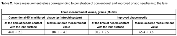
The experimental study was performed under operating room conditions at the mean ambient temperature of 22.5 ±0.4 оC. It was found that the force measurement at the time of direct contact of the conventional needle with the anterior lens surface was 44.0 ± 2.3 g. Further penetration of the phaco needle into the nucleus was characterized by plastic deformation of the nucleus with development of multiple radial cracks followed by destruction of the lens substance. The maximum force measurement for the conventional needle was 104.1 ± 4.3 g and corresponded to achieving the densest layers of the lens nucleus. The force measurement at the time of direct contact of the improved phaco needle with the anterior lens surface was 30.2 ± 2.5 g. This is mainly because the needle contact with flat surfaces oriented perpendicular to needle motion vector is practically excluded, thus reducing the total pressure on the lens. The maximum force measurement which corresponded to achieving the densest layers of the lens nucleus by the improved needle was 65.4 ± 3.6 g (Table 2).
Conclusion In the in vitro experimental study, it was established that the mean force measurement value at the time of direct contact of the improved phaco needle with the anterior lens surface and penetration of central lens layers by the needle was 30.2 ± 2.5 g and 65.4 ± 3.6 g, respectively. The mean force measurement value for the improved phaco needle was 1.4 times lower than that for the conventional needle. Therefore, the use of the improved needle for the phaco of a high-density nucleus is reasonable and makes it possible to penetrate deeply into the lens and divide the lens into fragments in a prompter and more efficacious manner than with the use of the conventional phaco needle. References 1. Kopaeva VG, Andreev IuV, Belikov AV et al. [Laser extraction of brown cataracts with an Nd-YAG 1.44 mcm laser]. Vestn Oftalmol. 2002 Jan-Feb;118(1):22-6. Russian 2. Korosteleva NF, Marchenkova TE. [Importance of biomicroscopy in the determination of density of cataract before phacoemulsification]. Vestn Oftalmol. 1989 Nov-Dec;105(6):43-5. Russian 3. Korosteleva NF, Nersesov IuE, Shalygin GF. [A method to determine nuclear hardness]. Oftalmokhir. 1990;1:42-5. Russian 4. Czygan G, Hartung C. Mechanical testing of isolated senile human eye lens nuclei. Med Eng Phys. 1996; 18(5): 345-9. 5. Heyworth P, Thompson GM, Tabandeh H. et al. The relationship between clinical classification of cataract and lens hardness. Eye (Lond). 1993;7 (Pt 6):726-30. 6. Tabandeh H., Thompson GM, Heyworth P. Lens hardness in mature cataracts. Eye (Lond). 1994;8 (Pt 4):453-8. 7. Avetisov KS. [New approaches to investigation of crystalline lens based on combined ultrasound technique]. [Cand. Sc. Thesis]. Moscow: Russian State Medical University; 2011. 128 p. Russian 8. Gritsenko YaA, Dmitriev SK, Kovalchuk AG et al. [An improved method for determining the density characteristics of the lens in patients with age-related cataract by ultrasound B-scan]. Oftalmol Zh. 2015;1:96-102. 9. Gritsenko YaA, Dmitriev SK, Pasyechnikova NV. [Improved needle for phacoemulsification of high-density nuclei]. Oftalmol Zh. 2015;1 Russian
|