J.ophthalmol.(Ukraine).2016;1:48-50.
|
https://doi.org/10.31288/oftalmolzh201614850 The role of activity indicators of redox enzymes in evaluation of cornea and conjunctiva condition in experimental hypothyroidism G.I. Drozhzhina, Dr. Sc. (Med), Prof M.I. Pavlovsky, PhD student SI "The Filatov Institute of Eye Diseases and Tissue Therapy of NAMS of Ukraine" Odessa, Ukraine Introduction. Thyroid diseases rank the second place after diabetes mellitus by disease incidence. In recent years, a large number of hypothyroidism studies have showed structural and functional abnormalities in eye tissues. Purpose. To analyze the activity of lactate dehydrogenase and malate dehydrogenase in tears, cornea and conjunctiva in experimental hypothyroidism. Material and Methods. Experimental studies were performed on rats. All the animals were divided into three groups: I – control group (14 rats), II – study group (14 rats), animals with the initial stage of hypothyroidism, and III – study group (14 rats), animals with the pronounced stage of hypothyroidism. In the tissues of the cornea, conjunctiva, and lacrimal fluid, the activity of lactate dehydrogenase and malate dehydrogenase was determined. Results. The data obtained on the reduction of the activity of lactate dehydrogenase and malate dehydrogenase evidence the deterioration of the intensity of the redox processes in tissues of the anterior eye in experimental hypothyroidism. Conclusions. In experimental hypothyroidism, the activity of LDH and MDH is reduced in the cornea by 29.2% and 23.7%, and by 34.7% and 28.7% in the conjunctiva, respectively. A significant increase in the activity of redox enzymes in the lacrimal fluid was revealed in the course of the pronounced hypothyroidism stage (LDH – by 36.3%, MDH – by 24.6%). At that, the degree of change in LDH activity depends on the stage of hypothyroidism. Key words: hypothyroidism, cornea, conjunctiva, lacrimal fluid, lactate dehydrogenase, malate dehydrogenase Introduction Thyroid diseases rank the second place after diabetes mellitus by disease incidence. In recent years, a large number of hypothyroidism (HT) studies have showed structural and functional abnormalities in eye tissues [4, 6, 8, 10]. Due to these papers, it became known that thyroid hormones level reduction is accompanied by metabolic, functional and structural changes in various organs and tissues, including the eye [6, 8]. At the same time, the absence of the literature data on the state of bioenergetics processes in the tissues of the eye in HT should be noted. Undoubtedly, pathogenetic mechanisms of HT-affected vision organ disorders have not been adequately studied yet, constituting an obstacle to the improvement of diagnostics and treatment methods. Proceeding from the aforesaid, it seems important to study redox processes in the lacrimal fluid and tissues of the anterior eye in experimental hypothyroidism. Objective: To analyze the activity of lactate dehydrogenase and malate dehydrogenase in the lacrimal fluid, cornea and conjunctiva in experimental hypothyroidism. Material and Methods Male albino Wistar rats weighing 190-210 g were used in the study, 42 animals in total. Requirements on biomedical researches on animals adopted by the Council for International Organizations of Medical Sciences (2012) were considered in the research performance. All the animals were divided into three groups: I – control group (14 rats), II – study group (14 rats), animals with the initial stage of hypothyroidism, and III – study group (14 rats), animals with the pronounced stage of hypothyroidism. Hypothyroidism was induced by using Tiamazol antithyroid medication, which was administered to the II and III group animals with drinking water (500 mg/l). Initial HT stage simulation was conducted within four weeks [9]. Pronounced HT stage simulation took place within 10 weeks of the medication administration [7]. Animals were withdrawn from the experiment by using ethyl ether excessive anesthesia. In the cornea, conjunctiva and lacrimal fluid tissues, LDH and MDH activity was determined. LDH activity determination is based on measuring the speed of enzymatic oxidation of the reduced NAD in the process of pyruvic acid recovery by the decrease of optical density of the investigated solution at a wavelength of 340 nm. The coefficient of variation of the method is 4.8% [5]. Malate dehydrogenase activity determination is based on measuring the oxidized NAD reduction speed in the process of malate oxidization by the optical density of the investigated solution increasing at a wavelength of 340 nm. The coefficient of variation of the method is 4.0% [5]. The results obtained were subjected to the appropriate statistical analysis using SPSS 11.0 package [1]. Results and Discussion LDH and MDH activity indicators in the anterior eye tissues and the lacrimal fluid under HT progression are shown in Table 1.
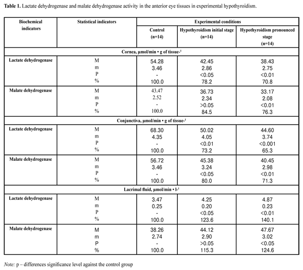
As it can be seen from the presented data, the activity of LDH in the cornea of animals at the initial stage of HT decreased to 78.2%; at the pronounced stage of HT, the lactate dehydrogenase indicators decreased to 70.8% as compared to the control group. The activity of MDH in the cornea of animals at the initial stage of HT was decreased to 84.5% comparing with the normal value; at the pronounced stage of HT, the malate dehydrogenase indicators decreased to 76.8%. At the initial stage of HT, the activity of LDH in the cornea of animals was decreased to 73.2%; at the pronounced stage of HT, lactate dehydrogenase indicators decreased to 65.3%. At the initial stage of HT, the activity of MDH in the conjunctiva of animals was decreased to 80.0%, at the pronounced stage of HT – to 76.8% comparing to the controls. In the lacrimal fluid of animals at the initial stage of HT, the activity of LDH increased up to 123.6%, and at the pronounced stage of HT, it increased up to 140.1% against the indicators of the control group. In conditions of pronounced HT stage, the activity of MDH in the lacrimal fluid increased up to 124.6% comparing to the control group. The data obtained on the reduction of the activity of lactate dehydrogenase and malate dehydrogenase in cornea and conjunctiva tissues indicate the deterioration of redox processes intensity in the tissues of the anterior eye in experimental hypothyroidism. At the same time, an increase of the activity of studied dehydrogenases in the lacrimal fluid can be considered as a result of the destruction of cells and subcellular structures, primarily in the cornea with hypothyroidism. This finding is consistent with research data of J. A. Petrovich and N. A. Terehina, pointing out that LDH activity determination in the lacrimal fluid is a sensitive method to detect even minor damages of the corneal epithelium [2, 3].
Conclusions 1. In experimental hypothyroidism, the activity of LDH and MDH was reduced in the cornea by 29.2% and 23.7%, and by 34.7% and 28.7% in the conjunctiva, respectively. 2. A significant increase in the activity of redox enzymes in the lacrimal fluid was revealed in the course of the pronounced hypothyroidism stage (LDH – by 36.3%, MDH – by 24.6%). Wherein, the degree of change of lactate dehydrogenase activity was determined by the hypothyroidism stage.
References 1. Nasledov A. [SPSS computer analysis of data in psychology and social studies]. St.-Petersburg: Piter Publ. 2005:416. Russian. 2. Petrovioch YA, Terekhina NA. [Biochemistry of tear and its changes in case of pathematoloy]. Voprosy med. khimii. 1990(36):13-18. Russian. 3. Terekhina NA, Petrovioch YA, Goldfeld NG. [Prediction of keratitis relapses via activity test of dehydrogenase of lacrimal fluid]. Zhurnal oftalmologii. 1988(5):42-44. Russian. 4. Babu K, Jayaraaj IA, Prabhaka J. Effect of Abnormal thyroid hormone changes in lipid peroxidation and Antioxidant imbalance in Hypothyroid and Hyperthyroid patients. Int. J Biol. Med. Res. 2011;2(4):1122–1126. 5. Bergmeyer HU. Methoden der enzymatischen Analyse. Berlin. 1986:2220. 6. Bruno AN, Diniz GP, Ricachenevsky FK. Hypo-and hyperthyroidism affect the ATP, ADP and AMP hydrolysis in rat hippocampal and cortical slices. Neurosci Res. 2005;52(1):61-68. 7. Dias AC, M?dulo CM, Jorge AG, Braz AM.Influence of thyroid hormone on thyroid hormone receptor beta-1 expression and lacrimal gland and ocular surface morphology. Invest. Ophthalmol Vis. Sci. 2012;52(7):3038-3042. 8. Gatzioufas Z, Panos GD, Brugnolli E, Hafezi E.Corneal topographical and biomechanical variations associated with hypothyroidism. J Refract Surg. 2014;30(2):78-79. 9. Ortiz-Butron R, Pacheco-Rosado J. Mild thyroid hormones deficiency modifies benzodiazepine and mu-opioid receptor binding in rats. Neuropharmacology. 2013;54(1):111-116. 10. Plummer CE, Specht A, Gelatt KN. Ocular manifestations of endocrine disease. Compend. Contin. Educ. Vet. 2013;31(12):733-743.
|