J.ophthalmol.(Ukraine).2015;6:17-22.
|
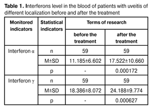
https://doi.org/10.31288/oftalmolzh201561722 Change of interferons ? and ? levels in the blood of patients with uveitis as a result of combination therapy with the use of cycloferon, decametoxine electrophoresis and microwave therapy N.V. Konovalova Chaibi Abderrahim State Institution "The Filatov Institute of Eye Diseases and Tissue Therapy of the National Academy of Medical Sciences of Ukraine'' Odessa (Ukraine) Key words: uveitis, medicamentous therapy, cycloferon, decametoxine electrophoresis, microwave therapy, interferons ? and ?, blood plasma. Introduction. Searching for new effective pathogenetic approaches to the uveitis treatment is both clinically and socially relevant, considering the pathology high prevalence and disease-induced disability. Objective. To determine the peculiarities of interferons ? and ? expression in the blood of patients with uveitis as a result of combination therapy with the use of cycloferon, decametoxine electrophoresis and microwave therapy. Materials and methods. Patients with endogenous uveitis received a combination therapy with the use of cycloferon, decametoxine electrophoresis and microwave therapy (26 persons). Control groups: cycloferon and decametoxine electrophoresis (20 persons) and cycloferon and microwave therapy (13 persons). The level of interferons ? and ? was measured in the blood plasma and the ophthalmological examination was carried out before and after the treatment. Parametric and nonparametric criteria, as well as correlation analysis, were used in statistical data processing. Results. A significant increase of interferons ? and ? concentration was found as a result of the treatment of patients with uveitis. The increase degree does not depend on the focus of inflammation location and the nature of the clinical course. At the same time, the most significant increases are pronounced the most in severe uveitis. There was found a reliable correlation between the level of interferons before and after the treatment, as well as the clinical characteristics of the disease. The increase of interferons ? and ? level was determined in various clinical forms of uveitis. The most pronounced increase of interferons ? and ? level was detected as a result of decametoxine electrophoresis and microwave therapy on the background of cycloferon administration. It amounts 56.7% and 33.4% respectively. Conclusions. The increase of interferons level was detected in the blood of patients after the treatment. The most significant increase occurred as a result of decametoxine electrophoresis and microwave therapy on the background of cycloferon administration. It may be considered as one of the treatment efficacy criteria. Introduction Endogenous uveitis is a widespread and difficult to treat disease that is often accompanied by complications and disability. Despite the numerous studies of the pathogenesis of this disease and the understanding of the underlying causes and mechanisms of its progression, the problem of the treatment efficacy remains unsolved [5, 6, 10]. Taking into account the known data on the role of the immune processes violations and the imbalance of pro- and anti-inflammatory cytokines in uveitis progression, the need for the use of immunotropic medications in this disease treatment is beyond doubt [2, 14]. However, when choosing medications the particularities of the immune status of the patient should be taken into account. Diagnostic significance of the cytokines level evaluation lies in ascertaining the fact of the disease focusing (the increase or the decrease of the concentration) and the severity of these changes in the patient with a particular disease. Although cytokines are local mediators, in this connection it is expedient to measure their level in the respective tissues or biological fluids. Determination of cytokines level in the plasma is extremely important because it reflects the general state of the immune system and protective reactions development, characterizing the organism immunoreactivity [11, 12]. Data on the content of cytokines in the peripheral blood can not only characterize the clinical features of the disease, but also the dynamics of the pathological process and the presence (or probability) of exacerbations [13]. It is logical to assume that according to the changing of the cytokine profile one can judge the effectiveness and appropriateness of the therapy. In order to suppress the autoimmune reactions, it is necessary to use the immunosuppressive agents (corticosteroids, cytostatic agents, cyclosporine A, FK-506). And to eliminate the immunodeficiency disorders it is necessary to use the immunomodulating agents (interferon inducers, activine t, and levamisole). Wherein it should be remembered that unreasonable and/or excessive immunosuppression or immunostimulation increases the risk of post-uveitis complications [9, 15]. Optimal, in this context, is the use of immunomodulators (myelopid, imunofan, activine t, levamisole, poludanum, plasmapheresis, hemosorption) weakening the hyperreactivity and / or enhancing reduced immunological indices, that is, normalizing them [8]. Widespread use of new medications in the clinic, anyhow modifying the cytokine status of the organism assumes controlling their level during treatment in order to provide correct cytokine therapy. Based on the foregoing, the determination of cytokines level, as one of the critical immunopathogenesis factor, in the uveitis treatment dynamics will provide the quantitative data required to monitor the immunocorrective effect of used medications or methods. Objective. Determination of the peculiarities of interferons ? и ? expression in the blood of patients with uveitis as a result of combination therapy with the use of cycloferon, decametoxine electrophoresis and microwave therapy. Material and methods 61 persons were observed and treated against endogenous uveitis in the Department of Inflammatory Eye Pathology of the State Institution "The Filatov Institute of Eye Diseases and Tissue Therapy of the National Academy of Medical Sciences of Ukraine''. Their age was between 13 and 76 with an average of (38.1 ± 13.2), including 33 women (54.1%) and 28 (45.9%) men. Distribution of the patients considering the etiological factors was presented in details previously [7]. 17 persons among the patients had anterior uveitis (including 8 with acute iridocyclitis and 9 with chronic iridocyclitis). 41 patients had posterior uveitis (including 19 persons with focal chorioretinitis and 14 with disseminated chorioretinitis and 1 with retinovaskulitis and 7 with optic neuritis). In one case panuveitis was diagnosed. Patients were examined within standard ophthalmic research protocol, including visometry, tonometry, ophthalmoscopy, visual field. For the possibility to analyze the efficacy of the proposed combination therapy the patients were divided into three groups: 1st group (20 persons) - patients received cycloferon and decametoxine by electrophoresis; 2nd group (13 persons) - patients received cycloferon and microwave therapy; 3rd group (26 persons) - patients received cycloferon, decametoxine by electrophoresis and microwave therapy. Cycloferon (''POLYSAN STPF'' LLC) was injected intramuscularly by 500 mg, daily scheme (1, 2, 4, 6, 8, 11, 14, 17, 20, 23), 10 injections, total dose 5.0 g. Decametoxine (CJSC "Darnitsa" Pharmaceutical Company) was used as 0.02% aqueous solution according to the standard procedure of electrophoresis. During transorbital electrophoresis, an active electrode in the form of a tray was poured with 2-3 ml of 2% calcium chloride solution and then with 1 ml of decametoxine. An indifferent electrode was in the neck area. During intranasal electrophoresis, an active electrode had the form of nasal turunda. In both cases, an anode is positive; the current is increased in stages from 0.3-0.5-0.8 mA to 1 mA, duration of the procedure 3-5-10 minutes. The gap between the two types of electrophoresis - 2 hours. Microwave therapy was carried out using "Luch-2'' apparatus though the contact method in which the electrode is located on the skin of closed eyelids. D'Arsonval currents frequency - 50000 - 100000 fluctuations per second. The exposure is 5-10 minutes and the overall course is 10 sessions. In the blood plasma of 59 persons the level of ? and ? interferons was determined before and after the treatment by means of solid-phase immune-enzyme analysis (kits of reagents made by "Vector-Best", Novosibirsk, Russia). Units of interferon content (pg/mL) [3, 4]. The obtained data was statistically processed with the use of Statistica 7.0 software and the parametric Student criterion for pair wise comparison of two groups and preliminary assessment of distribution normality, as well as Spearman rank correlation criterion to identify the link between studied parameters [1]. Results and their discussion The results of our previous research [7] have revealed a significant correlation between the clinical characteristics and the features of the pathological process progression and the initial level of interferons ? and ? in the blood of patients with uveitis. Thus, it has been shown that the content of these cytokines is significantly lower in the posterior uveitis and in chronic course of the disease with a high degree of reliability. The focusing and the intensity of the expression of the abovementioned interferons determination as a result of the treatment in general for the whole group has shown a significant increase in concentration both interferon ?, and interferon ?: by respectively 1.56 and 1.3 times (p = 0.0001 and 0.0006) (Table 1).
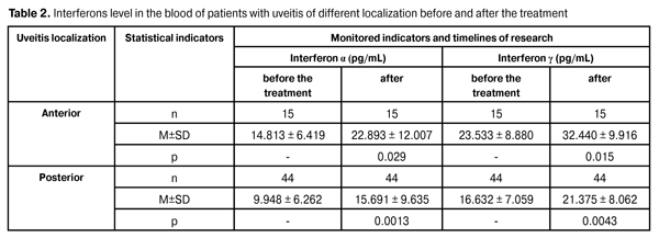
An analysis of the relevant data, taking into account the localization of the pathological process (Table 2) revealed a similar focusing of changes, which in all cases were reliably significant. It was also shown that the severity of changes does not depend on the location of the disease (interferon ? level after the treatment of the anterior and posterior uveitis has increased in 1.55 and 1.58 times (p = 0.029), and interferon ? - in 1.38 and 1.29 times, respectively) (p = 0.015), i.e., the increase of ? interferon is more pronounced.
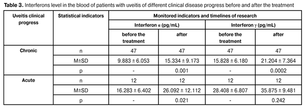
Uveitis clinical course particularities (chronic or acute) also do not impact the dynamics indicators of interferon content in the blood of patients as a result of the treatment. In both cases there is interferon ? concentration increase in 1.5 times, and interferon ? in 1.34 and 1.26 times respectively (p = 0.0002). These changes are valid except for the data on the increase of interferon ? after the treatment in the group of patients with acute uveitis as a tendency (Table 3).
The most pronounced increase of interferons after the treatment occurs in severe uveitis (Table 4). Thus, the content of interferon ? increases nearly twofold (in 1.88 times), and interferon ? in 1.39 times. However, these changes are not reliable, which can be explained by the small number of observations in the group (n = 5 before the treatment and 6 after the treatment) and sufficiently large spread of individual values in different patients. In persons with an average severity of uveitis the differences between the levels of studied interferons before and after the treatment are reliably relevant and are 56 and 30%, respectively (p = 0.0004 and 0.0013).
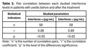
Pair correlation analysis data has shown a strong correlation between the levels of studied interferons in patients with uveitis before and after the treatment: correlation coefficient is 0.88 and 0.89 for interferon ? and interferon ?, respectively (Table 5).
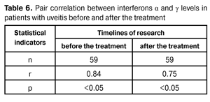
The same strong reliable correlation was found between the values characterizing the level of interferons ? and ? before the treatment (r = 0.84), as well as their values, achieved as a result of the treatment (r = 0.75) (Table 6).
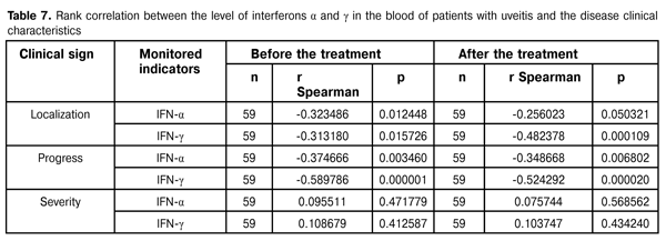
The data shown in Table 7 indicates the presence of a rank correlation between the clinical characteristics of the disease (uveitis localization and its progression peculiarities), and the level of interferons before and after the treatment.
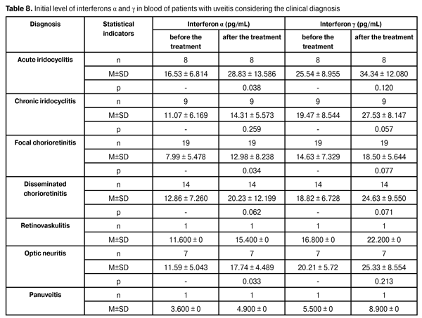
Characterizing the dynamics of interferons expression as a result of the treatment, taking into account the clinical diagnosis, it should be noted that the fact of these cytokines level increasing takes place in every known uveitis form, but the changes are reliably significant only for interferon ? in acute iridocyclitis, focal chorioretinitis, optic neuritis, constituting respectively 174, 162 and 153% relative to the corresponding data before the treatment. In other cases, the character of the changes can be classified as a tendency to increase (Table 8).
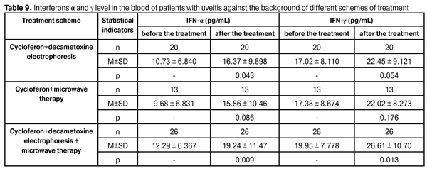
Analyzing the data that characterizes the level of interferons in the blood of patients with uveitis given the diagnosis (acute and chronic iridocyclitis, disseminated chorioretinitis, retinal vasculitis, optic neuritis, panuveitis) using the Kruskal-Wallis criterion, there was no significant correlation found between the average concentrations of interferon ? both before (?2=5.8402, df=6, p=0.441), and after the treatment (?2=7.468, df=6, p=0.280). Similar data was obtained for interferon ?. Connection between the average values of this cytokine according to the Kruskal-Wallis criterion before the treatment and after the treatment is not reliable (?2= 9.230, df = 6, p = 0.161 and ?2= 9.392, df = 6, p = 0.153 respectively). The results obtained with this approach to the statistical experimental data array processing can be explained by the different number of observations in each group, including the isolated retinal vasculitis and panuveitis cases among the observed patients. To evaluate the electiveness of the proposed combination therapy as the comparison we have representatively marked the groups of patients in which on the top of already administered cycloferon therapy decametoxine electrophoresis, as well as microwave therapy were applied. The obtained results (Table 9) show a positive effect which was evaluated by the degree of interferons level increase in all groups. When comparing the data characterizing the interferons content in the blood after the treatment, it was found that the most pronounced increase in the level of interferons occurs in all the observed groups, but in patients who received the combination therapy (cycloferon + decametoxine electrophoresis + microwave therapy), these changes were reliably significant and constituted 56.7% for interferon ? and 33.4% for interferon ?, respectively, with p = 0.009 and 0.013. In general the results obtained allow the following conclusions. Conclusions 1. The significant increase in the concentration of interferons ? and ? resulting from the treatment in the whole for the group was shown - in 1.56 and 1.3 times, respectively. 2. The increase degree of the studied interferons level after the treatment does not depend on the localization of the inflammatory focus, however, it was more pronounced for interferon ?, which level has increased in anterior and posterior uveitis in 1.55 and 1.58 times, whereas interferon ? - in 1.38 and 1.29 times. 3. The character of the clinical course of uveitis (acute or chronic) does not determine the dynamics of the content of interferons in the blood of patients after the treatment (in both cases, concentration interferon-? increases reliably by half, interferon ? in 1.34 and 1.26 times, respectively). 4. The most pronounced interferons increase after the treatment occurs in severe uveitis: interferon ? - in 1.88 times, interferon ? - in 1.39 times; in persons with average severity of uveitis, these differences are respectively 56 and 30%. 5. In patients with uveitis a strong correlation between the level of each researched interferons before and after the treatment was found (the correlation coefficient for interferon ? and ? is respectively 0.88 and 0.89), between the interferons level indicators ? and ? before the treatment (r = 0.84), as well as their values, achieved as a result of the treatment (r = 0.75). 6. The rank correlation was established between the clinical characteristics of the disease (uveitis localization and its progress features) and interferons level before and after the treatment. 7. Interferons ? and ? level increase was noted in different clinical forms of uveitis, being reliably significant in acute iridocyclitis, focal chorioretinitis and optic neuritis for interferon ?, constituting 174, 162 and 153 % regarding the data before the treatment.
8. Interferons level increase in the blood of patients after the treatment was found. It was pronounced the most as a result of decametoxine electrophoresis and microwave therapy under pressure of cycloferon therapy. The changes are reliably significant, constituting for interferon ? and ? 56.7 and 33.4%. References 1. Glanz S. [Biomedical statistics]. Glanz S.: translation from English. M.: Practice. 1998:459. Russian. 2. Yershov FI, Narovlyansky MV, Mezentseva MV[Early cytokine responses in viral infections]. Tsytokiny i vospalenie. 2004;13(1):3-7. Russian. 3. Instruction for using the kit of reagents for quantitative determination of human alpha interferon in biological fluids and human culture mediums. ZAO "Vector-Best''. Novosibirsk; 30.05.08:22. Russian. 4. Instruction for using the kit of reagents for quantitative determination of human gamma interferon in biological fluids and human culture mediums. ZAO "Vector-Best''. Novosibirsk; 30.05.08:22. Russian. 5. Katargina LA, Khvatova AV, Krichevskaya N and others. [Clinical variations and immunological features of rheumatoid uveitis in children of different age]. Vestnik. oftalmol. 2001;117(4):30-33. Russian. 6. Kovalchuk LV, Gankovskaya LV, Rubakov EI. [System of cytokines]. Moscow; 2000:324. Russian. 7. Konovalova NV, Chaibi Abderrahim. [Level of interferon ? and ? in the blood of patients with uveitis with different clinical characteristics and course of the disease]. Oftalmol. Zh. 2015;3:24-29. Russian. 8. Neverova EA. [Pathogenetic substantiation of ozone use in endogenous uveitis]. Dissertation of Candidate of Medical Sciences 14.00.16 Patologicheskaya fiziologiya. 2007; Saransk:116. Russian. 9. Ospelnikova TP, Mironova TV, Poloskov VV. Gharib FYu, Ershov FI. [Effect of interferon inducers on the cytokine profile]. Tsytokiny i vospalenie. 2014;13(1):37-40. Russian. 10. Roit A, Brostoff J, Mail D.[Immunology]. Transl. from English. M.: Mir. 2000:581. Russian. 11. Ryabicheva TG, Varaksin NA, Timofeev NV, M. Rukavishnikov MY. [Determination of cytokines by ELISA of JSC "Vector-Best"]. Informatsionnyi biulleten Novosti "Vector Best". 2004; 4(34). http://www.vectorbest. ru/nvb/n34/st34_4.htm. Russian. 12. Simbirtsev A. [Cytokines is a new system of regulation of body defense reactions]. Tsytokiny i vospalenie. 2002;11(1):9-16. Russian. 13. Khaitov RM, Pinegin BV, Yarilin AA. [Guidelines of clinical immunology. Diagnosis of diseases of the immune system: a guide for doctors]. M.: GEOTAR Media. 2009:352. Russian. 14. Abbas AK, Lichtman AH, Pillai S. Cellular and molecular immunology. 6th ed: Philadelphia; 2009:581. 15. Mahon CR, Tice D. Clinical laboratory immunology. New Jersey: Upper Side River. 2006:325.
|