J.ophthalmol.(Ukraine).2015;6:3-5.
|
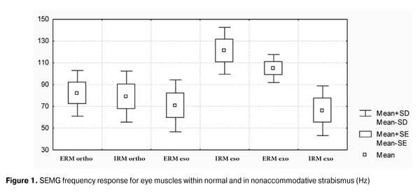
https://doi.org/10.31288/oftalmolzh2015635 Features of bioelectric potentials of the extraocular horizontal rectus muscles in concomitant esotropia and exotropia I.M. Boichuk, Doctor of Medical Sciences V.P. Mazur Filatov Eye Disease and Tissue Therapy Institute, the NAMS of Ukraine Odessa (Ukraine) Introduction. There is no objective method currently available for assessing the extraocular muscles functional status, which could be considered definitely effective and convenient for the extensive use in ophthalmological practice. Also, there is no comparative analysis of bioelectric potentials of the extraocular horizontal rectus muscles in concomitant esotropia and exotropia. The purpose of the study was to investigate the functional status of bioelectric potentials of the extraocular horizontal rectus muscles in concomitant esotropia and exotropia using surface electromyography (SEMG). Materials and methods. There were 18 children under follow-up (12.9 ± SD3.2) years of age, of which 12 (24 eyes) had concomitant esotropia and 6 (12 eyes) with concomitant exotropia, and healthy children (28) of the same age. Horizontal muscle functional status was determined by computer myograph M-TEST-2. Results. The frequency in esotropia of bioelectric potentials of the internal rectus muscle is much higher than the frequency of the extraocular rectus muscle (128 Hz ± 19.35 and 60.9 ± 16.7 Hz, respectively, p = 0.0003). The frequency in exotropia of bioelectric potentials of the extraocular rectus muscles was in average higher than the frequency of the intraocular rectus muscle (109 ± 9.9 Hz and 55.0 ± 1.57, respectively, p = 0.004). The frequency response when compared in healthy children showed no differences between the internal and external line (63.0 ± 19.9 Hz and 63.7 ± 21.1 Hz, p> 0.05). Conclusions. 1. The results suggest that the children with concomitant strabismus have functional disorders of the horizontal rectus muscles, as evidenced by SEMG data. 2. It was established that the bioelectric potentials frequency of the intraocular rectus muscle in esotropia and extraocular rectus muscle in exotropia is higher than the frequency of the relevant muscles in the healthy children. 3. The bioelectric potential of the intraocular and extraocular rectus muscles in the healthy children does not differ, and in children with concomitant esotropia and exotropia differs significantly. Introduction Strabismus is one of the visual organ severe functional and cosmetic defects, which is an external manifestation of a deep sensory and motor abnormality. Oculomotor apraxia, according to the literature, is found in 0.5-7.1% of children [1]. Complex ophthalmoneurological studies [1-3, 12, 13] revealed that the vast majority of nonaccommodative strabismus cases are of paretic nature and one of the congenital or transferred in the early stages of child's development neurological impairment symptoms. Oculomotor adromia (OM) causes a change in their function and leads to the motor imbalance, which manifests in a great variety of strabismus clinical forms [1, 5, 8, 9, 10, 14]. In cases when there is a clinical paresis cure the strabismus remains as a result of secondary contralateral synergist and ipsilateral antagonist hyperfunction. The interest in the literature increased in recent years for the oculomotor muscles function study. Various aspects of electromyography (EMG) are described with greater detail in review articles and monographs [7, 8, 17, 16, 15, 18]. However, EMG using concentric needle electrodes for an objective investigation of the oculomotor muscles function failed due to the invasive techniques to obtain extensive use in clinical practice, which sharply restricts its use in pediatric patients [4, 18]. In order to investigate OM function some authors [11] suggest using electrooculography. This study results depend on the patient's psychophysiological condition, eye movement’s amplitude and velocity, as well as retina functional integrity, and therefore the technique is unreliable. All of the above served as an impetus to the development of non-invasive surface electromyography (SEMG) method for the eye extraocular horizontal muscles to assess their functional status. Modern multichannel electromyographs allow recording bioelectric activity of muscles, but they have no special electrodes for the eye muscles bioelectrical activity recording. Therefore, given the size of the eye extraocular rectus muscles and limb attachment distance in children, two silver and platinum electrodes were modified with a diameter of 5 mm and spacing of 6 mm, and SEMG technique proposed [6]. The possibility of this study is confirmed in literature [19], which shows that theevoked potentials obtained from the surface electrodes placed on the eyeball in place of the oculomotor muscles attachment are derived by the muscle responses. The aim of the study was to evaluate the functional status of the extraocular horizontal rectus muscles in concomitant esotropia and exotropia using surface electromyography (SEMG). Material and methods There were 18 children under follow-up (12.9 ± SD3.2) years of age, of which 12 (24 eyes) had concomitant esotropia and 6 (12 eyes) with concomitant exotropia, and healthy children (28) of the same age. The visual acuity in patients with the correction was in average 0.85 ± 0.15. The horizontal muscles functional status was assessed using computer electromyograph M-TEST-2 [6]. To remove the bioelectric potentials at SEMG two silver and platinum electrodes were used with a diameter of 5 mm and spacing of 6 mm. The active electrode was placed above the rectus muscle venter at maximum eyes abduction to the opposite direction, and passive one – above the limb. The study was conducted after local anesthetic instilling in the following sequence: extraocular rectus muscle, intraocular rectus muscle of the right eye, at a maximum levoduction and abduction to the right, and then extraocular and intraocular rectus muscle of the left eye at the maximum levoduction and abduction to the right. Measuring the interference EMG parameters: total frequency of the muscles electrical activity; maximum and average signal amplitude was conducted in triplicate for each muscle. Results The study results of the surface electromyography bioelectric potentials frequency response are shown in Figure.1. Data analysis of the frequency response for SEMG bioelectric potential of the horizontal rectus muscles in the concomitant esotropia showed that the average frequency of the intraocular rectus muscle frequency is much higher that of the extraocular rectus muscles – (128 ± 19.35) Hz and (60.9 ± 16.7) Hz, respectively, p = 0.0003. In concomitant exotropia the total frequency of the extraocular rectus muscle electrical activity is on average higher than that of the extraocular rectus muscle – (109 ± 9.9) Hz and (55.0 ± 1.57) Hz, respectively, p = 0.004. When comparing the frequency response in healthy children no differences between the internal and external line were found (63.0 ± 19.9) Hz (21.1 ± 63.7) Hz, P> 0.05. It was found that the bioelectric potentials frequency of the intraocular rectus muscle in patients with esotropia, and the external line with exotropia is significantly higher than in healthy patients (128 ± 19.35) Hz compared to (63.0 ± 19.9) Hz, p = 0.0001 (109 ± 9.9) Hz (21.1 ± 63.7) Hz, P = 0.0001, respectively.
Conclusions 1. The results obtained suggest that the children with concomitant strabismus have functional disorders of the horizontal rectus muscles, as evidenced by SEMG data. 2. It was established that the bioelectric potentials frequency of the intraocular rectus muscle in esotropia and extraocular rectus muscle in exotropia is higher than the frequency of the relevant muscles in the healthy children. 3. The bioelectric potential of the intraocular and extraocular rectus muscles in the healthy children doesn’t differ, and in children with concomitant esotropia and exotropia differs significantly. References 1. Avetisov ES [Guidelines of pediatric pediatric ophthalmology]. In: Kovalevsky EI, Khvatova AV. M. Meditsina, 1987:185. Russian. 2. Avetisov ES [Concomitant strabismus]. M .: Meditsina. 1977. Russian. 3. Gromakina EV [Pathogenetic aspects of strabismus in children with perinatal pathology: author's thesis for the degree of Doctor of Medical Sciences]. Krasnoyarsk. 2002. Russian. 4. Gecht BM [Theoretical and clinical electromyography]. L. Nauka. 1988. Russian. 5. Kashchenko TP [New possibilities of diagnostics and treatment of oculomotor pathology]. In: IX Congress of Russian Ophthalmologists: thes. of reports. Moscow, 2010. Russian. 6. Boychuk IM, Mazur VP [A new method of surface electromyography of direct eye muscles in children]. Oftalmol. Zh. 2014;3:15-18. Russian. 7. Kryzhanovsky GN, Pozdnyakov OM, Polgar AA [Pathology synaptic apparatus muscles]. M. Meditsina, 1974. Russian. 8. Plisov IL [Experience of paralytic strabismus treatment by hemodenervation of extraocular muscles]. In: сoll. of scient. papers. Ophthalmology of the Black Sea. Krasnodar, 2006. Russian. 9. Rozemblyum YZ, Chernyshov SG [Diagnosis and comprehensive treatment of diplopia muscle origin; method. recommendations]. M. 2004. Russian. 10. Rozemblyum YZ [Rehabilitation of patients with diplopia: Method. recommendations]. Ministry of Health of the RSFSR. [Ed. Rozemblyum YZ, Kashchenko TP], 1988. Russian. 11. Semyonovskaya EN, Khvatova AV [Electrooculography in strabismus]. In: Concomitant strabismus and amblyopia. The researchers note. M. 1962. Russian. 12. Smolyaninova IL [Paretic strabismus in children: author. of dis. ... Dr. med. Sciences]. M. 1972. Russian. 13. Shaytor VM [Long-term effects of perinatal damage to the nervous system in children: author's thesis for the degree of Doctor of Medical Sciences]. SPb. 2008. Russian. 14. Adler’s Physiology of the Eye. Mosby Year Book. 1991:641- 707. 15. Breinin GM and Moidaver J. Electromyography of human extra ocular muscles. In: Arch. Opht. 1955;54:206. 16. Desmedt JE. New Developments in Electromyography and Clinical Neurophysiology. Basel. Karger. 1973:1-3. 17. Engel WK, Brooke MH, Nelson PG. Histochemical studies of denervated or tenotomized cat muscle illustrating difficulties in relating experimental animal conditions to human neuromuscular diseases. In: Ann. N.Y. Acad. Sci. 1966. 18. Lennerstrand G. What can eye muscle studies tell us about strabismus? In: Transactions 28th Meeting ESA. Bergen. Norway. 2003:65-75. 19. Sasaki T, Suzuki K, Matsumoto M. et al. Origins of surface potentials evoked by electrical stimulation of oculomotor nerves: are they related to electrooculographic or electromyographic events. J Neurosurgery. 2002. Oct;97(4):941-944.
|