J.ophthalmol.(Ukraine).2015;5:52-57.
|
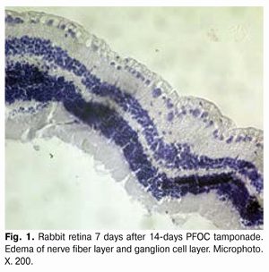
https://doi.org/10.31288/oftalmolzh201555257 Structural changes of the rabbit retina after 14-days tamponade of the vitreal cavity with perfluoro-organic compounds Zhmurik D.V. 1, PhD in Medical Sciences Vit V.V. 2, prof., Doctor of Medical Sciences Dumbrova N.E. 2, prof., Doctor of Medical Sciences Molchaniuk N.I. 2, PhD in Biological Sciences Miliienko M.V. 1, MD 1 Kiev Municipal Clinical Ophtalmological Hospital “The Center of Eye Microsurgery”; Kiev (Ukraine) 2 State Institution “The Filatov Institute of Eye Diseases and Tissue Therapy of the National Academy of Medical Sciences of Ukraine”; Odessa (Ukraine) E-mail: vizus@ukr.net Introduction. Perfluoro-organic compounds (PFOC) have valuable qualities for vitreoretinal surgery and their application for short-term tamponade could expand indications for preoperative treatment and improve its results. However, there is no unambiguous opinion concerning the mechanical action of PFOC on ultrastructural structure of retina. The purpose of the study: To study the influence of 14-days PFOC tamponade on the structure of the rabbit in experiment; to compare the action of PFOC, saline solution, light and heavy silicone oil in dynamics after the tamponade completion. Material and methods. Light and electro- microscopic study was carried out on 36 rabbits (72 eyes). All animals underwent back closed subtotal vitrectomy followed by 14-days PFOC tamponade (right eye), light and heavy silicone and saline solution (left eye). The study was conducted 7, 14 and 30 days after the completion of 14-days tamponade. Results. PFOC, light and heavy silicone cause similar changes in the ultrastructure of the studied retina elements. These are mainly hydropic changes of smooth endoplasmic reticulum and mitochondria of the retinal pigment epithelium (RPE) cells, mitochondria of inner segments of photoreceptor cells, ganglion cells and Muller cells. At the same time, these cells also show the signs of compensatory-restorative processes, which is reflected in increasing of protein synthesis and energy producing activity that contributes to normalization of the revealed damage of ultrastructure. It should be noted that PFOC and light silicone have particularly similar effect on the retina, while heavy silicone causes more pronounced reaction at subcellular level. Introduction of the saline solution during the first periods of observation leads to the light reactive hydropic changes, mainly in PRE cells. Conclusion. Revealed hydropic changes in the retina after the completion of 14-days tamponade with the used substances are classified as reactive, not damaging changes and are reversible. By the end of the periods of observation the ultrastructure of the retina practically restores. Therefore PFOC can be considered as an alternative for a short-term tamponade. Key words: light optical and ultrastructural changes, retina, perfluoro-organic compounds (perfluorocarbonliquid), light and heavy silicone oil Introduction Usage of the substances with a high specific gravity for the short-term postoperative tamponade during the surgical treatment of rhegmatogenous (complicated by anterior proliferative vitreoretinopathy), and far advanced traction diabetic retinal detachments (RD) could expand indications for surgical treatment and improve not only anatomical but also functional results of the treatment. The specific gravity of perfluoro-organic compounds (PFOC) is two times bigger than this of the water and thousand times bigger than this of the air. PFOC are chemically and metabolically stable, transparent and have a low viscosity. For the first time they were presented in the medicine in 1966 [5]. From the beginning of the eighties, liquid perfluorocarbons due to its gas transmission function have been used as blood substitutes (perfluoran). The first experience of intravitreal use of PFOC belongs to Haidt and colleagues. They noted the absence of gross lesions of the retina, lens and cornea within the period of observation up to three months after the surgery, allowing the further use of PFOC in vitreoretinal surgery [8] .However, the attitude of vitreoretinal surgeons to the short-term tamponade (7-14 days) of vitreous cavity with PFOC is ambivalent. Mechanical damaging effect of PFOC remains an open question [2, 3, 4]. Most researchers report about the maximally safe period of PFOC tamponade during two weeks [1, 12]. Therefore, it would be important to compare the mechanical action of PFOS, heavy fluoride-containing (specific gravity 1.02-1.06 g/cm3) and light silicone oil (viscosity 5700 cSt, specific gravity 0.971-0.975 g/cm3). Considering that heavy and light silicones are routinely used for the postoperative tamponade of vitreous cavity, PFOC with different specific gravity 1.54 to 1.94 g/cm3 (perfluoro-n-octane - 1.76 g/cm3, perfluorotributylamine - 1.89 g/cm3, pefluordekalin - 1.94 g/cm3, and others) are used in clinical practice. For experimental studies it is advisable to use PFOC with high specific gravity, as absence of damage during their usage indirectly indicates the safety of other types of PFOC with less specific gravity. Also, it should be noted that rabbit always remains in the same position and for studying the effect of heavy silicone and PFOC during the electron-microscopic study (EMR) the lower segments of the retina should be separated; for studying the impact of light silicone oil it should be done with the upper segments. In experimental works devoted to this issue, the authors studied the effect of PFOC on the retina of experimental animals with help of ERG, light and electron microscopy, performed without completion of tamponade, or in different periods after removing of PFOC from the vitreous cavity with one fixed term of tamponade [2, 3, 4, 6, 7, 10, 13]. In our opinion, it makes impossible to assess the degree of reversibility of retinal changes after a short-term PFOC tamponade and surgical trauma. Objective of the study is to study of the impact of the short-term PFOC tamponade (14 days) on the structure of rabbit retina in the experiment; to compare the effects of PFOC, saline solution, light and heavy silicone oil in dynamics through performance of light-optical studies (LOS) and electron-microscopic studies (EMS) at different times points after the tamponade completion (7, 14, 30 days). Material and methods The experimental study was conducted on 36 male Chinchilla rabbits (72 eyes) with weight of (3.5 ± 0.5) kg, at age of (6.5 ± 0.5) months. The duration of the tamponade with PFOC was 14 days. All animals underwent LOS and EMS of the retina at different time points, in 7, 14 and 30 days after the completion of vitreous cavity tamponade with PFOC, silicone and saline solution. In all cases, the second eye (left) was the controlled one. The controlled eyes underwent the tamponade with light silicone oil with viscosity of 5700 cSt, heavy silicone oil and saline solution. All surgical interventions, as well as removal of the animals from the experiment were performed in accordance with the “Rules of the Treatment with Laboratory Animals” [9]. Methods of surgical intervention Anesthesia: thiopental sodium solution in a dose of 2 mg/kg intramuscularly, 0.5% proxymetacanie solution epibulbarly. Mydriasis: 1 drop of 1% atropine sulfate and 2.5% phenylephrine epibulbarly. 0.3 % ofloxacin solution epibulbarly before the surgery. Back closed subtotal vitrectomy (BCSV) was performed under control of surgical microscope OPTON: OpMi-8 by KFE-01 "MEDA-NN" device (frequency up to 1200 beats/min, aspiration of 150 mm Hg) using 20, 23 and 25G instruments. 1.5 ml of PFOC – perfluorodekalin was injected into the cavity of the right eye (16 rabbits). 1-1.5 ml of light silicone oil with viscosity of 5700 cSt (16 rabbits), saline solution (16 rabbits) or 1-1.5 ml of heavy silicone oil (16 rabbits) was injected into the cavity of the left eye (control injection). After completion of the vitrectomy, 0.3% ofloxacin ointment was put in the conjunctival cavity. Completion of the tamponade was carried out after performing the preparation described above. Withdrawal of PFOC, heavy and light silicone was performed under control of the surgical microscope OPTON: OpMi-8 by RFE-01 "MEDA-NN" device (aspiration of 150 mm Hg). In the eyes which underwent tamponade with saline solution the solution was replaced. For LOS the selected retina of enucleated eyeball was fixed in 10% neutral formalin, and subsequently encapsulated in paraffin. The sections were stained with hematoxylin and eosin. The light-optical study was performed using a microscope Jenamed 2. For EMS the fragments of retinal tissue (lower segments of the retina during the tamponade with PFOC and heavy silicone oil and upper segments during the tamponade with light silicone oil with viscosity of 5700 cSt) were fixed in 2.5% glutaraldehyde solution on phosphate buffer with pH – 7.4 followed by additional fixation with 1% osmic acid solution with the same pH of the buffer solution. After that, the samples were dehydrated in alcohols of increasing strength. The material was soaked and encapsulated in Epon-Araldite mixture. Then ultrathin sections were contrasted using the method of Reynolds [11]. The material was studied using the electron microscope PEM-100 - 01. Results and their discussion Structural changes of retina at 14-days tamponade with PFOC. LOS showed that after 14 days of PFOC tamponade in all cases there was observed edema of the nerve fiber layer and the ganglion cell layer (Fig. 1).
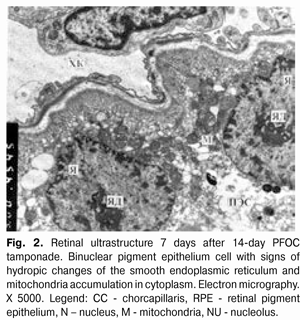
During EMS, 7 days after the completion of PFOC tamponade, the ultrastructure of choriocapillaris (CC) remains unchanged. The retinal pigment epithelium (RPE) cells are partially fragmented; sometimes there is some destruction and disintegration of individual cellular elements. However, these cells have a lot of normal organelles and there are some cells containing 2 nuclei, i.e. along with the events of destruction, mainly of smooth endoplasmic reticulum (SER) membranes and large mitochondria, there are signs of activation of intracellular activity (Fig. 2).
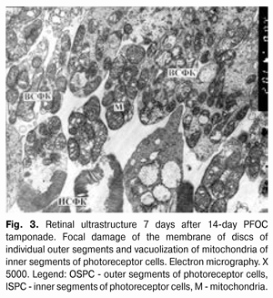
The pathology of disks of the outer segments (OS) of photoreceptor cells (PC) and vacuolization of mitochondria in the inner segment (IS) of PC is observed in individual PC (Fig. 3).
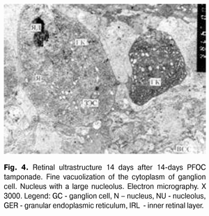
The inner reticular layer shows hydropic changes of the structures. In the layer of ganglion cells (GC) there was a fine vacuolization of Muller cells (MUC) processes and edema of GC mitochondria. 14 days after PFOC tamponade completion the CC are mainly extremely expanded. Contents of the lumen of microvessels are fine-grained, with moderate electron density. RPE cells contain regular organelles, basal and apical regions are well expressed. Cytoplasm shows a small vacuolization of mitochondria and SER elements. SER membranes are loose, somewhere fragmented. In the PC layer some cells have intracellular edema of inner segments and pathology of mitochondria. Area of PC nuclei is not changed. Neuronal cells of the inner segments of the retina are unchanged. There are pronounced hydropic changes of the inner retinal layer. GC layer cells contain nuclei with large nucleoli and cytoplasm with large clusters of elements of granular endoplasmic reticulum (GER) and small vacuolated mitochondria. MUC processes around the cell of GC layer contain small vacuoles (Fig. 4).
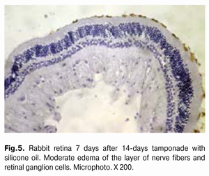
30 days after PFOC tamponade completion CC are expanded. Lumen content is regular. RPE cells are unchanged as well as have a number of pathological changes: vacuolization, and sometimes destruction of SER elements. However, some cells contain two nuclei, large clusters of mitochondria with signs of activity. Apical microvilli of RPE cells are destroyed in some areas. In PC there are elements of edema in the inner segments and individual damages of membrane structures in the outer segments. There are hydropic changes observed in the inner retinal layer. Large cells in GC layer have a large number of organelles involved in protein synthesis activity, as well as mitochondria and others. MUC processes around GC have very fine vacuolization. Structural retina changes during 14-days tamponade with silicone oil. LOS showed structural changes of the retina that were almost similar to those observed during the application of PFOC and consisted in moderate edema in the layers of nerve fibers and ganglion cells (Fig. 5).
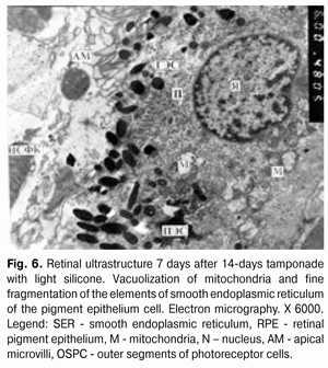
7 days after completion of the tamponade with light silicone oil, EMS showed signs of alteration of the structure of RPE cells: fine fragmentation of SER membrane elements and vacuolization of mitochondria (Fig. 6).
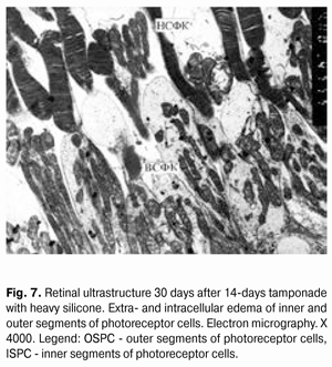
There is a destruction of disks of outer segments and edema of inner segments in PC. Other retinal cellular elements have no visible changes. Hydropic changes in the structures of the inner retinal layer are observed. Small hydropic changes also occur in GC layer - both in neuronal and in glial ones. 14 days after the tamponade with light silicone oil the remains of hydropic changes still can be found in RPE cells. There is a destruction of OS discs in individual PC. No visible changes in the retinal layers were established. Small hydropic changes were found in GC and MUC. 30 days after the tamponade with light silicone oil reactive changes in ultrastructure of individual nervous structures of the retina are practically defined. The received data are difficult to be differentiated from the artifacts brought during the experiment. Structural retina changes during 14-days tamponade with heavy silicone oil. During EMS it was established that 7-14 days after the completion of the tamponade with heavy silicone oil the CC lumen is expanded. There is a vacuolization of SER observed in RPE cells. There is a loosening of the area of outer and inner PC segments. Signs of hydropic changes of the outer and inner layers of the retina and vacuolization of GC cytoplasm and MUC processes in GC layer. 30 days after the completion of the tamponade there is a fragmentation of elements of SER and vacuolization of mitochondria in cytoplasm of RPE cells. There is a dilution of OS and ISPC in PC area. A part of the outer segments has damaged disks. Edema is observed in the inner segments but with remaining ultrastructure of mitochondria (Fig. 7).
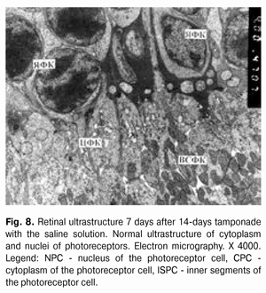
Hydropic changes are observed in the structures of the outer and inner retinal layers. There are small hydropic changes in GC, while at the same time vacuolization of the cytoplasm is observed in MUC processes located at GC and the internal limiting membrane. However, the described changes in ultrastructure of the retina cover a much smaller range of nervous elements of the retina than it was observed 14 days after the tamponade completion. Structural changes of the retina during 14-days tamponade with saline solution. 7 days after 14-days tamponade with saline solution, LOS have found a mild edema in the retinal layers, which has been reduced by the 30th day of observation and the structure of the retina looks practically unchanged (Fig. 8).
7 days after, EMS has found small hydropic changes of SER elements of RPE cytoplasm and separation of OS PC. A number of studied cellular elements of subsequent retinal layers showed the enlightenment of cytoplasmic matrix.
In 14 and 30 days after the tamponade with the saline solution the ultrastructural condition of the retinal elements is normal. Conclusion The performed study highlights the same type of qualitative and quantitative changes in the structure of the retina – both when using PFOC and silicone oil. Changes are reduced to edema of the inner layers of the retina, with different severity, which can be observed on the 7th day and remains throughout the entire period of observation. It should be noted that edema of the retina is detected during the application of the saline solution as well, but it is negligible and is localized predominantly in the layer of the nerve fibers. All three types of the studied impact on the retina have specific similar effect on the ultrastructure of the studied retinal elements. These are, basically, hydropic changes in the smooth endoplasmic reticulum cells of the retinal pigment epithelium, pigment epithelium mitochondria, inner segments of photoreceptor cells, ganglion cells, and Muller cells. At the same time compensatory-restorative processes appear in these cells that help to normalize their ultrastructure. Differences in the effect of PFOC and silicone on the retina mainly consisted in the dynamics of the observed ultrastructural changes. In addition, PFOC and light silicone have particularly similar effect on the retina, while heavy silicone causes more pronounced reaction at the subcellular level. The findings suggest that changes in the elements of the retina are reversible and can be normalized; and do not have destructive effect while using these compounds for the tamponade of vitreous cavity during 14 days. Control injection of the saline solution during all periods of the study causes mild hydropic reactive changes, mostly in the retinal pigment epithelium cells. Series of works are devoted to the research of the effect of PFOC on the retinal tissue, but the attitude of vitreoretinal surgeons to the tamponade of vitreous cavity with PFOC is ambivalent. Some researchers have reported about development of irreversible atrophic changes 48-hours [6], 2-weeks [4] after PFOC tamponade. The difference with our data can be explained by unequal conditions of experimental studies. Some authors have used gas compression of the vitreous body, which could cause additional damaging effect on the retinal ultrastructure. Absence of any significant changes after more than 14-day tamponade is reported in other experimental studies; it corresponds to our data [7]. In all the above mentioned works, saline solution has been used as a control. In this experimental study we compared the effect of PFOC on the structure of the retina, not only with the effect of saline solution, but also with widely used plugging substances, such as light and heavy silicone, and found no significant difference in the effect of these substances on the structure of the retina. Since the effect of 14-day tamponade with PFOC on ultrastructural structure of the retina is comparable with a standard widely used plugging agent – light and heavy silicone – it can be considered as alternative during the performance of the short-term tamponade. Literature 1.Takhchidi KP, Kostin OA. [Specifics of the surgery of tractional retinal detachment at proliferative diabetic retinopathy]. Perfluoro-organic compounds in Biology and Medicine. Sb Nauch Tr. Pushchino. 1999:192-194. Russian. 2.Shkvorchenko DO. [Complex surgical treatment of the retinal detachment, complicated by giant gaps and separation from dentate line with application of liquid perfluoro-organic compounds]. Dis…PhD in Med. Sci. 14.00.08. Moscow. 1995: 132. Russian. 3.Shkvorchenko DO, Kashtan OV, Makarov KN, Ronkina TI. [Experimental clinical application of vitreopress for the short-term postoperative tamponade in vitreoretinal surgery]. Perfluoro-organic compounds in Biology and Medicine. Sb Nauch Tr. Pushchino. 1999: 186-192. Russian. 4.Chang S, Sparrow JR, Iwamoto T, Gershbein A, Ross R, Ortiz R. Experimental studies of tolerance to intravitreal perfluoronoctane liquid. Retina. 1991; 11: 367-374. 5.Clark LC Jr, Gollan F. Survival of mammals breathing organic liquids equilibrated with oxygen at atmospheric pressure. Science. 1966; 152: 1755-1756. 6.Devin F, Jourdan T, Saracco JB, Lucciani A. Experimental tolerability of perfluorodecalin in prolonged intraocular tamponade. J Fr Ophtalmol. 1995; 18: 268–274. 7.Flores-Aguilar M, Munguia D, Loeb E, Crapotta JA, Vuong C, Shakiba S, Bergeron-Lynn G, Wiley CA, Weers J, Freeman WR. Intraocular tolerance of perfluorooctylbromide (perflubron). Retina. 1995; 15: 3-13. 8.Haidt SJ, Clark LC, Ginsberg J. Liquid perfluorocarbon replacement of the eye. Invest. Ophthalmol.VisSci. 1982; 22: 233. 9.Norman H J. Requirements of bioethics of the Helsinki declaration about ethical regulation of medical researches. Chronicle of the World Organization of Healthcare. 1985; 39 (3): 3-9 10.Orzalesi N, Migliavacca L, Bottoni F, Miglior S. Experimental short-term tolerance to perfluorodecalin in the rabbit eye: a histopathological study. Curr Eye Res. 1998; 17: 828–835. 11.Reynoldes ES. The use of lead citrate at high pH an electronopaque stain in electron microscopy. I. Cell Biol. 17: 208-212. 12.Sirimaharaj M, Balachandran C, Chan WC, Hunyor AP, Chang AA, Gregory-Roberts J, Hunyor AB, Playfair TJ. [Vitrectomy with short term postoperative tamponade using perfluorocarbonliquid for giant retinal tears]. Br J Ophthalmol. 2005; 89: 1176-1179. 13.Terauchi H, Okinami S, Kozaki Z, Tanihara H, Nagata M, Segawa Y. Experimental study on the effects of a replacement of the vitreous body with perfluorotributylamine on the rabbit eye. Nihon Ganka Gakkai Zasshi. 1989; 93: 294-301.
|